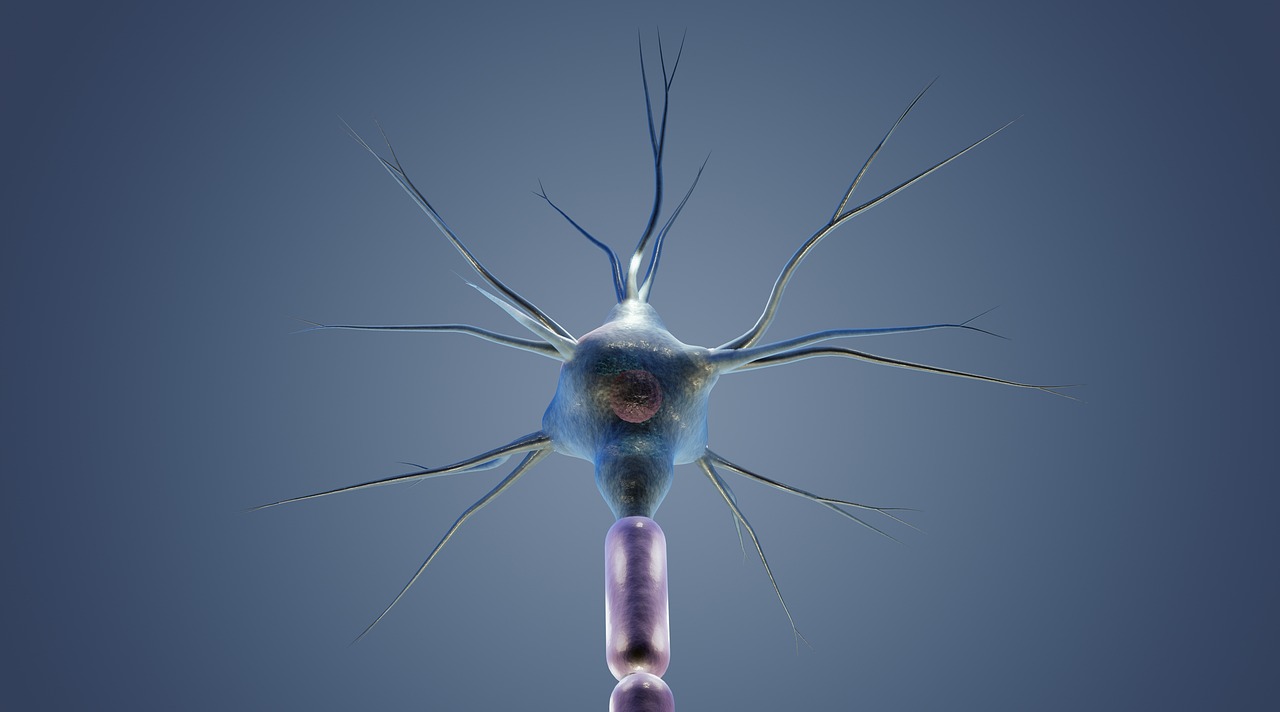
New evidence suggests that the cells responsible for communication in the brain may have different structures in children with autism. Researchers at the Del Monte Institute for Neuroscience at the University of Rochester have found that neuron density in certain areas of the brain varies in children with autism compared to the general population.
“We have spent many years describing the broad characteristics of brain regions, such as thickness, volume, and curvature,” said Zachary Christensen, MD/PhD candidate at the University of Rochester School of Medicine and Dentistry, and the first author of the paper published today in Autism Research. “However, newer techniques in the field of neuroimaging for characterizing cells using MRI reveal new levels of complexity throughout development.”
Imaging provides new insight into brain development
Researchers analyzed brain imaging data from over 11,000 children aged 9-11. They compared brain scans of 142 children with autism to those of the general population and found lower neuron density in certain regions of the cerebral cortex. These regions are associated with memory, learning, reasoning, and problem-solving. On the other hand, they also found increased neuron density in the amygdala, a region responsible for emotions. The researchers compared the brain scans of children with autism not only to those without any neurodevelopmental diagnosis but also to a large group of children diagnosed with common psychiatric disorders like ADHD and anxiety. The results indicated that these differences are specific to autism.
“People diagnosed with autism often have to cope with other conditions such as anxiety, depression, and ADHD. These findings indicate that we now have a new set of measurements that show promise in identifying individuals with autism,” Christensen explained. “If we can reliably and easily identify unique differences in neuron structure in those with autism, it opens up opportunities to understand how autism develops. These measures could also be used to identify individuals with autism who may benefit from more targeted therapeutic interventions.”
Technology leverages what we know about the inner workings of the brain and autism
The advancement of technology has greatly improved the ability of investigators to observe precise details in neuronal structure. In the past, researchers could only observe structural differences in neural populations after death. The imaging data for this research was obtained from the Adolescent Brain Cognitive Development (ABCD) study database, which is the largest long-term study of brain development and child health. The University of Rochester is one of 21 national sites collecting data for this study, which began in 2015 and has significantly enhanced our understanding of adolescent brain health and development.