Making small changes in your life can and will lead to positive outcomes in the future. Becoming more engaged in life and not sweating the small stuff. To have the courage to step through the doors life presents and then maybe to go on a journey you never imagined. Diagnosed with Multiple Sclerosis in 1995, Rob has taken on the challenge of a TEDx talk the way he deals with pretty much everything.
A study of more than 22,000 people with multiple sclerosis (MS) has discovered the first genetic variant associated with faster disease progression, which can rob patients of their mobility and independence over time. The work resulted from a large international collaboration of more than 70 institutions worldwide, led by researchers from UCSF (USA) and the University of Cambridge (UK). About 3 million people live with multiple sclerosis or MS, but the real number may be higher. Women are more likely to develop MS than men. When diagnosed, the immune system attacks the central nervous system, causing symptoms that range from numbness to disability. Although scientists know MS is associated with genetic risks, MS is not an inherited disease, and doctors have struggled to understand why some cases progress faster than others. This latest study combed through 13,000 patients’ DNA and found a gene that is linked to the onset of severe disability. Sergio Baranzini is a Neurology Professor at the University of California San Francisco. He is the study’s lead author, and he joins us live from San Francisco to discuss the latest updates.
Immigrants to Canada who have spent a greater proportion of their lives in Canada have a greater risk of developing multiple sclerosis (MS) than people who have spent a smaller proportion of their lives there, according to a new study. The study does not prove that an increased proportion of life in Canada causes MS; it only shows an association.
“Other studies have shown that immigrants tend to have better health than long-term residents, which is thought to be because healthy people are more likely to choose to immigrate,” said study author Manav V. Vyasy. “We wanted to see if the lower risk of MS declines over time as people adopt some of the unhealthy lifestyles of their new country or are exposed to other environmental factors that increase their risk.”
The study involved 1.5 million immigrants who arrived in Canada between 1985 and 2003 and were covered by health insurance for at least two years with no diagnosis of MS. The people were then followed through 2016.
During that time, 934 people were diagnosed with MS, a rate of 0.44 cases per 100,000 person-years. Based on previous research, the overall rate of MS in Canada is estimated to be 15 to 17 cases per 100,000 person-years. Person years represent the number of people in the study and the amount of time each person spends.
The person’s age at arrival in Canada and the amount of time since they immigrated determined the proportion of life spent in Canada. Overall, people have spent an average of 20% of their lives in Canada.
Researchers found that people who had spent 70% of their lives in Canada were 38% more likely to develop MS than people who had spent 20% of their lives there. This result took into account other factors that could affect the risk of MS, such as sex, age and other health conditions.
The researchers did not find any differences between men and women or between people belonging to one of Canada’s immigration classes: family, refugee, or economic.
“Our data did not include information on various environmental factors associated with MS, but our theories include that this increase in the risk of MS over time may be due to lifestyle factors such as higher rates of smoking and changes in diet, environmental factors such as sunlight exposure and biological factors such as the composition of the gut microbiome that have been previously associated with an increased risk of MS,” Vyas said. “Some immigrants may be more susceptible to these risk factors due to social determinants of health such as income, education, neighbourhood and access to nutritious food.”
Actor and advocate Selma Blair tells Meet the Press Moderator Kristen Welker that she lives “in pain every day” after her multiple sclerosis went into remission and opens up about her treatment.
The new technique could lead to more advanced treatments for multiple sclerosis.
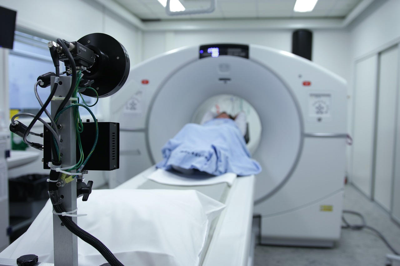
A new study from Brigham and Women’s Hospital, a founding member of the Mass General Brigham healthcare system, suggests that positron emission tomography (PET) brain scans could reveal hidden inflammation in patients with multiple sclerosis (MS) who are being treated with highly effective treatments. The findings were published in Clinical Nuclear Medicine.
“One of the perplexing challenges for clinicians treating patients with MS is after a certain amount of time, patients continue to get worse while their MRIs don’t change,” said lead author Tarun Singhal, MD, MBBS, an associate professor of Neurology in the Brigham’s Department of Neurology and director of the PET Imaging Program in the Ann Romney Center for Neurologic Diseases. “This is a new approach that is potentially going to be very helpful for the field, for research, and hopefully for clinical use.”
Singhal collaborated with others in the Brigham Multiple Sclerosis Center and the Ann Romney Center. The study started when Singhal noticed that patients treated with the most effective MS treatments were experiencing worsening symptoms. The team has worked for the past eight years on developing an approach of imaging cells called microglia. Microglia are immune cells in the brain that are thought to have a role in MS disease progression but cannot be seen by a routine MRI. The team developed a technique called F18 PBR 06 PET imaging. It involves the injection of a tracer, or dye, that binds to the microglia cells.
Rohit Bakshi, MD, of the Department of Neurology and a co-author on the paper, said increased microglial activity means more atrophy of grey matter in the brain.
“This can affect cognition, movement, fine motor skills, and other aspects of their life,” Bakshi said.
In their paper, the authors describe the term “smouldering” inflammation. Just as a smouldering fire can burn slowly without smoke or flame, smouldering inflammation may linger in patients with MS, driving disease progression and symptoms, even when it cannot be assessed on MRI.
The newly published study involved performing PET scans on 22 people with MS and eight healthy controls. Researchers measured the glial activity load on the PET scans, a new measure developed in Singhal’s lab where lab members looked at the level of smouldering inflammation from microglia in MS patients. They compared those scans to patients’ disability and fatigue levels and not only found that PET scans could show hidden inflammation caused by microglia, but the damage to patients’ brains correlated with the disability and fatigue levels they were experiencing. The researchers were also able to better classify patients with MS between high-efficacy and low-efficacy treatments. Those with low-efficacy treatments had more abnormalities on their PET scans, suggesting more microglial cell activation. Those using high-efficacy treatments had a lower degree of PET abnormality than those on no or low-efficacy treatments but still had an abnormal increase of microglial activation compared to healthy people, suggesting that while high-efficacy treatments helped to reduce neuroinflammation, there was residual inflammation despite treatment, which could account for future worsening and progression independent of relapse activity (PIRA) in these MS patients.
“Our therapies are excellent in that we’ve improved MS patients’ lives,” Bakshi said. “There’s no doubt about that, but we’re still not at the finish line.”
One limitation to the study is the initial group was small. The authors note that PET scans can also be expensive and expose patients to some level of radiation, whereas MRIs do not. Singhal said that radiation could be reduced because of the long half-life and the requirement for a lower administered dose of the F18 PBR06 tracer. The tracer also produces better imaging characteristics than previously used tracers with shorter half-lives.
Bakshi said despite the limitations, the study shines an important light on the power of PET scanning, specifically to find microglial activation.
“This study tells us something new about the disease and may be giving us an important clue as to what is driving disease progression in patients,” he said.
Singhal said before the technique can be used routinely in a clinical setting, it must be validated on a larger sample size. Other longer half-life PET tracers have been approved by the FDA for clinical use, for example, amyloid PET tracers for studying Alzheimer’s disease. If approved, [F-18]PBR06 could also be used to personalize and predict a patient’s treatment course in MS and other brain diseases. However, the authors note that even before approval, [F-18]PBR06 can help advance drug development and perform multicentric clinical trials.
“It’s very exciting that our novel approach worked and correlated so strongly with clinical measures we assessed,” he said. “It means our approach is relevant clinically.”
We use cookies on our website to give you the most relevant experience by remembering your preferences and repeat visits. By clicking “Accept”, you consent to the use of ALL the cookies.
This website uses cookies to improve your experience while you navigate through the website. Out of these, the cookies that are categorized as necessary are stored on your browser as they are essential for the working of basic functionalities of the website. We also use third-party cookies that help us analyze and understand how you use this website. These cookies will be stored in your browser only with your consent. You also have the option to opt-out of these cookies. But opting out of some of these cookies may affect your browsing experience.
Necessary cookies are absolutely essential for the website to function properly. This category only includes cookies that ensures basic functionalities and security features of the website. These cookies do not store any personal information.
Any cookies that may not be particularly necessary for the website to function and is used specifically to collect user personal data via analytics, ads, other embedded contents are termed as non-necessary cookies. It is mandatory to procure user consent prior to running these cookies on your website.