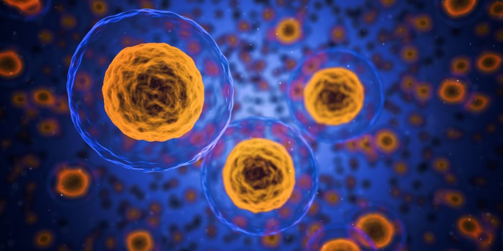
Targeting type of B cell could reduce lupus disease, study suggests

Targeting type of B cell could reduce lupus disease, study suggests


A group of University of Pittsburgh researchers has given new meaning to “knowing your ABCs.” In a new study, published in the Journal of Experimental Medicine, the team showed that a type of immune cell called age-associated B cells, or ABCs, are important drivers of lupus and that targeting these cells in a mouse model reduced disease, pointing the way to new therapies.

The immune system usually does a good job of discriminating between healthy body tissues and potential threats such as bacterial or viral infections. When exposed to a pathogen, B cells make antibodies, which help the body to recognize and eliminate the threat. The immune system then returns to less-active surveillance mode, while retaining a long-lived “memory” of prior infection.

However, in autoimmune disorders such as lupus, B cells become abnormally activated and begin attacking the patient’s organs, including the kidneys, lungs and skin.

“A hallmark of lupus is high levels of autoantibodies that bind to a patient’s own DNA or RNA,” said lead author Dr. Kevin Nickerson, research assistant professor in Pitt’s Department of Immunology.


“Because these genetic materials are never eliminated from the body, the immune system is continually reactivated, leading to inflammation that, over time, causes a great deal of damage to the patient’s body.”

According to Nickerson, existing therapies that broadly target B cells can be effective in some lupus patients, but because they also impair B cells that produce infection-fighting antibodies, these treatments can compromise immunity, suggesting a need for more narrowly focused approaches.

In the new study, Nickerson, senior author Dr. Mark Shlomchik, distinguished professor of immunology at Pitt, and their team set out to better understand the role of ABCs in lupus. Nickerson explains the study’s findings and what they could mean for the future of lupus treatments.

What are age-associated B cells?

KN: Age-associated B cells, or ABCs, are a sub-category of B cell that were first identified as being more frequent in older individuals than younger individuals. It quickly became apparent to researchers that these cells are also found in greater numbers in patients with certain autoimmune diseases, including lupus, and during certain chronic infections such as malaria and HIV. These and other findings led researchers to propose that ABCs are a type of immune memory cell that form during some types of chronic inflammatory immune responses. They might accumulate very slowly over a healthy individual’s entire lifetime, or much more rapidly in a younger patient with lupus.

What motivated this study?

KN: Previous research had shown that higher numbers of ABCs in a lupus patient’s blood often correlated with the severity of their symptoms, particularly lupus nephritis (kidney disease). ABCs were thought to be autoimmune memory B cells that develop into cells that make lupus autoantibodies. In this study, we set out to test whether ABCs were indeed a driver of lupus disease. Using a preclinical mouse model of lupus, we examined ABCs in great detail, studied their relationship to other types of B cells and genetically depleted these cells right after they are formed to see what effect they have on disease.

What were your main findings?

KN: Our most important finding was that reducing the number of ABCs by eliminating them as soon as they form slowed or reduced disease progression in the kidneys in mice, directly demonstrating that ABCs drive disease in this lupus model.  We also found that ABCs are a more diverse subset of B cells than previously known, which could suggest that they have different functions or that multiple pathways are involved in their formation.

We also demonstrated that ABCs are indeed a direct precursor to the cells that make lupus autoantibodies. Although they resemble anti-pathogen memory B cells in some respects, they seem to undergo continual cycles of reactivation, proliferation and differentiation. This makes sense because the DNA and RNA to which they respond are always present, so they can’t fully enter a resting state, unlike the memory response in B cells that recognize pathogens, which are eventually eliminated.

What are the implications of these findings and what do they tell you about potential therapies for lupus?

KN: Several lupus therapeutics in the clinic today aim to reduce B cell numbers by directly killing them or by blocking the factors necessary for their survival. However, these currently available therapies are nonspecific, meaning that they affect all B cells, whether those B cells are autoreactive or directed against pathogens. While depleting all B cells prevents lupus progression and organ damage, it significantly impairs a patient’s ability to respond to infections.

If ABCs are the pathogenic population of B cells in lupus, as our study suggests, then narrowly targeted therapies that focus on eliminating these “bad” cells, while sparing “good” B cells, could be beneficial. To do this, we need a more complete understanding of ABCs and where they come from to help design the best approaches. Our study contributes to this understanding.

What are the next steps for this research?

KN:  An important unresolved question is what makes ABCs so pathogenic? And how are they actually promoting disease? One part of that could be their role in making autoantibodies, but we have reason to believe that ABCs could also activate the T cell arm of the immune system in lupus. Autoreactive T cells, when activated, infiltrate organs and tissues from the bloodstream, causing injury by damaging cells and disrupting normal organ function. We want to know if ABCs are directly promoting this pathogenic process and if they are also found within the inflamed tissues at the sites of damage.  In addition, we want to develop methods to deplete ABCs during ongoing disease in a preclinical model to determine if and how that could slow down or treat disease.

Researchers identify gene mutations capable of regulating pain.

Gene mutation capable of regulating pain


Vanessa O. Zambelli and PhD candidate Beatriz Stein Neto. In a study involving mice, the scientists discovered that an avian variant of the TRPV1 receptor CREDIT Rafael Porto

Pain afflicts at least 1.5 billion people worldwide, and despite the availability of various painkilling drugs, not all forms of pain are treatable. Moreover, pain medications can have side-effects such as dependence and tolerance, especially in the case of morphine and other opioids. 

In search of novel painkillers, researchers at Butantan Institute’s Special Pain and Signaling Laboratory (LEDS) in São Paulo, Brazil, studied TRPV1, a sensory neuron receptor that captures noxious stimuli, including heat and the burning sensation conveyed by chili peppers, and discovered a potential pain insensitivity mutation in the gene that encodes this protein. They report their findings in an article published in the Journal of Clinical Investigation.

The study was supported by FAPESP and conducted in partnership with Stanford University and Emory University in the United States, and Münster University Hospital in Germany. The researchers analyzed a number of mutations in humans and also benefited from existing knowledge of birds, which unlike mammals have a TRPV1 receptor that is naturally resistant to noxious insults and even peppery food, yet can still perceive pain.

“There are more than 1,000 TRPV1 mutations in humans, and there’s nothing novel about trying to switch the receptor off in order to relieve pain, but these attempts haven’t been successful until now,” said Vanessa Olzon Zambelli, a researcher at LEDS and co-first author of the article. “First, many drugs resulting from this process interfere with body temperature regulation. Second, TRPV1 is an important channel for signaling heat, and completely altering its activity cancels out physiological pain, interfering with the sensation of burning heat, which has a protective function.”

The researchers began by exploring a genome database to compare the genetic sequences of avian and human TRPV1. Using a computational approach, they identified five avian mutations they believed to be linked to resistance to pain. Cryogenic electron microscopy (which does not require large sample sizes or crystallization and is therefore suited to the visualization of structures at near-atomic resolution) showed that the five avian mutations were located in K710, an amino acid residue believed to control gating (opening and closing) of the TRPV1 channel.

The mutations can also be present in humans, but they are very rare, so the researchers decided to find out what would happen if they were “transplanted” into mammals. When they tested these variants in genetically modified cells, they found that the function of the channel was indeed altered. Next, they used the CRISPR/Cas9 gene editing technique to create mice with the mutation K710N, which they had previously found to reduce the receptor’s reaction to capsaicin in cells. Capsaicin is the active principle in pepper.

The researchers did not observe nociceptive behavior (suggesting avoidance of pain) in mice with the K710N mutation injected with capsaicin and given peppery chicken feed, in contrast with the behavior of normal mice, which lifted their paws to avoid touching the capsaicin, presumably because even skin contact caused pain.

The mice with the K710N mutation also showed less hypersensitivity to nerve injury, while their response to noxious heat remained intact. Furthermore, blocking the K710 region in normal mice limited acute behavioral responses to noxious stimuli and returned pain hypersensitivity induced by nerve injury to baseline levels.

In addition to modulating pain, TRPV1 also plays an important role in protection against other stimuli. For example, recent evidence suggests that it serves in non-neuronal cells as an intracellular molecular sensor that protects against glucose-induced cellular stress or tissue ischemia. Additional tests performed as part of this study involving cardiomyocytes (heart muscle cells) insulted with hydrogen peroxide, high levels of glucose and a cerebral ischemia model confirmed the protective effect even with the mutation.

Translational analysis

The second part of the study consisted of an attempt to reduce the receptor’s function pharmacologically. To this end, the researchers developed a peptide, V1-cal, which acted selectively on the K710 region. Mice treated with V1-cal and given capsaicin displayed less nociceptive behavior and diminished release of neuropeptides leading to neurogenic inflammation and edema without altering temperature. Lastly, chronic pain also improved considerably.

“We now want to add value to this study by validating the results under best-practice laboratory conditions [required by regulatory agencies], identify other small molecules besides the peptide that can more easily be synthesized, conduct preclinical trials and, if these are successful, begin a clinical trial,” Zambelli said.

UC Davis study uncovers age-related brain differences in autistic individuals.

GABA cells

GABA cells in fuchsia, and vGLUT cells in red and yellow CREDITUC Regents

Genes involved in inflammation, immune response and neural connectivity behave differently in brains of autistic folks

A new study led by UC Davis MIND Institute researchers confirms that brain development in people with autism differs from those with typical neurodevelopment. According to the study published in PNAS, these differences are linked to genes involved in inflammation, immunity response and neural transmissions. They begin in childhood and evolve across the lifespan.

About one in 44 children in the U.S. has autism. Autistic individuals may behave, communicate and learn in ways that are different from neurotypical people. As they age, they often have challenges with social communication and interaction.

The researchers aimed to understand how neurons in the brain communicate and the interaction between age and autism. They studied the genetic differences in brain neurons in people with autism at different ages and compared them to those with neurotypical development.

Earlier studies have shown that certain brain regions mark early excess, followed by reductions in volume, connectivity, and cell densities of neurons as autistic folks age through adulthood.

“Initial excess and overconnectivity of neurons may make the brain more vulnerable to early aging and inflammation, which may lead to further changes in the brain structure and function,” said co-senior author Cynthia Schumann. Schumann is a professor of neuroscience in the Department of Psychiatry and Behavioral Sciences. She is affiliated with the UC Davis MIND Institute. “Understanding how the brain in a person with autism changes throughout life will provide opportunities for early intervention.”

Method

The researchers analyzed brain tissues from 27 deceased autistic individuals and 32 without autism. The age of these individuals ranged between 2 and 73 years.

The tissues were taken from the superior temporal gyrus (STG) region — an area in the brain responsible for sound and language processing and social perception.

The STG plays a critical role in integrating information. It helps provide meaning about our surroundings. Despite its importance, it remains relatively unexplored,” Schumann commented. “We wanted to understand how the molecular changes in this critical part of the brain are happening in autism.”

The team analyzed brain tissues as well as isolated neurons using laser capture microdissection techniques. They studied mRNA expression on a molecular level in the STG tissue and the isolated neurons. The mRNA translates the DNA code into instructions the cell machinery can recognize and use to make proteins for different body functions.

Main findings

Cynthia Schumann

Professor Cynthia Schumann in her laboratory with post-doctoral student Kari Hanson CREDIT UC Regents

The study identified 194 significantly different genes in the brains of people with autism. Of those genes, 143 produced more mRNA (upregulated) and 51 produced less (downregulated) in autistic brains than in typical ones.

The downregulated genes were mainly linked to brain connectivity. This may indicate that the neurons may not communicate as efficiently. Too much activity in the neurons may cause the brain to age faster in autistic individuals.

The study also found more mRNA for heat-shock proteins in autistic brains. These proteins respond to stress and activate immune response and inflammation.

Age-related brain differences between neurotypical and autistic people

The study identified 14 genes in bulk STG tissue that showed age-dependent differences between autistic and neurotypical individuals and three genes in isolated neurons. These genes were connected to synaptic as well as immunity and inflammation pathways.

For example, in typical brains, the expression of the HTRA2 gene is much higher before age 30 and decreases with age. In the STG neurons of people with autism, the expression levels of this gene begin lower and increase with age.

“Changes in HTRA2 have been implicated in neuronal cell loss and cell functions – such as proper protein folding, and reusing and recycling cell components,” explained co-senior author Boryana Stamova, associate professor in the Department of Neurology. She is also affiliated with the MIND Institute. “HTRA2’s role is vital for normal brain function.”

Broyana Stamova

Boryana Stamova is an associate professor at the Department of Neurology CREDIT UC Regents

The researchers also uncovered different inflammation patterns in autistic brain tissues. Several immune and inflammation-related genes were strongly upregulated, indicating immune dysfunction that may get worse with age.

The study pointed to an age-related decrease in the gene expression involved in Gamma-aminobutyric acid (GABA) synthesis. GABA is a chemical messenger that helps slow down the brain. It works as an inhibitory neurotransmitter.

“GABA is known for producing a dampening effect in controlling neuronal hyperactivity in anxiety and stress. Our study showed age-dependent alterations in genes involved in GABA signaling in brains of people with autism,” Stamova said.

The study found direct molecular-level evidence that insulin signaling was altered in the neurons of people with autism. It also noted significant similarities of mRNA expressions in the STG region between people with autism and those with Alzheimer’s disease. These expressions may be linked to increased likelihood of neurodegenerative and cognitive decline.

“The findings from our study are really important in understanding what is happening in the brains of people with autism. Identifying these changes over time gives us an opportunity to think about some interventions that might be more useful in certain periods,” Schumann said.

Credits

The study’s co-authors are Bradley Ander of the UC Davis MIND Institute and the Department of Neurology; Alicja Omanska of the UC Davis MIND Institute and the Department of Psychiatry and Behavioral Sciences; and Michael Gandal and Pan Zhang of UCLA.

Can the Mediterranean diet help people with Multiple Sclerosis preserve thinking skills?

Can Mediterranean diet help people with MS preserve thinking skills?


 People with multiple sclerosis (MS) who follow a Mediterranean diet may have a lower risk for problems with memory and thinking skills than those who do not follow the diet, according to a preliminary study released today, March 1, 2023, that will be presented at the American Academy of Neurology’s 75th Annual Meeting being held in person in Boston and live online from April 22-27, 2023.

The Mediterranean diet includes a high intake of vegetables, legumes, fruits, fish and healthy fats such as olive oil, and a low intake of dairy products, meats and saturated fatty acids.

“It’s exciting to see that we may be able to help people living with MS maintain better cognition by eating a Mediterranean diet,” said study author Ilana Katz Sand, MD, of the Icahn School of Medicine at Mount Sinai in New York, New York, and a member of the American Academy of Neurology. “Cognitive difficulties are very common in MS, and they often get worse over time, even with treatment with disease-modifying therapies. People living with MS are very interested in ways they can be proactive from a lifestyle perspective to help improve their outcomes.”

The study involved 563 people with MS. People completed a questionnaire to show how closely they followed the Mediterranean diet. They were assigned a score of zero to 14 based on their responses with higher scores given to those who more closely followed the diet.

Researchers then divided participants into four groups based on their diet scores, with the lowest group having scores of zero to four and the highest group having scores of nine or higher. 

Participants also took three tests assessing their thinking and memory skills. Cognitive impairment was defined as scoring less than the fifth percentile on two or three of the tests.

A total of 108 people, or 19%, had cognitive impairment.

The researchers found that people who more closely followed the Mediterranean diet had a 20% lower risk for cognitive impairment than people who did not follow the diet.

Among those in the lowest diet score group, 43 of 133 people, or 34%, had cognitive impairment compared to 13 of 103 people, or 13%, of people in the highest diet score group.

The relationship was stronger among people with progressive MS, where the disease steadily worsens, than among those with relapsing-remitting MS, where the disease flares up and then goes into periods of remission.

Importantly, Katz Sand noted, the results were the same when researchers rigorously adjusted for other factors that could affect the risk of cognitive impairment, such as socioeconomic status, smoking, body mass index, high blood pressure and exercise.

“Among health-related factors, the level of dietary alignment with the Mediterranean pattern was by far the strongest predictor of people’s cognitive scores and whether they met the study criteria for cognitive impairment,” Katz Sand said.

She noted that longer studies that follow people over time and well-designed interventional clinical trials are needed to confirm the results. A limitation of the study was that tests were taken only once.

Researchers bioengineer an endocrine pancreas for type 1 diabetes

Transplanting donor islet tissue to a bioengineered omentum—the fatty tissue that drapes from the stomach over the intestines—normalized blood glucose levels in nonhuman primates with type 1 diabetes.


In people with type 1 diabetes, the body’s immune system attacks and destroys insulin-producing β cells that control blood glucose levels and are part of a group of cells in the pancreas called pancreatic islets. In research published in Cell Reports Medicine, a team led by investigators at Massachusetts General Hospital (MGH), a founding member of Mass General Brigham, recently developed an efficient way to transplant pancreatic islets and demonstrated that the method can effectively reverse type 1 diabetes in nonhuman primates.

Pancreatic islet transplantation is a promising treatment approach for type 1 diabetes; however, current methods involving transplanting islets to the liver are inefficient and can result in the loss of as much as half of transplanted β cells due to immune attack. Also, the liver can only accommodate a limited volume of transplanted tissue. Scientists have wondered if an alternative site might provide a more hospitable environment and lead to better results. One promising site is the omentum, the fatty tissue that starts in the stomach and drapes over the intestines.

To optimize the omentum as a transplant site in an individual, investigators used topical recombinant thrombin (which stops bleeding), an enzyme, and the recipient’s own plasma to engineer a bio-degradable matrix by which donor islets are immobilized onto the omentum. When this strategy was used along with an immunosuppressive therapy to protect islets from immune attack, the method normalized blood glucose levels and restored glucose-responsive insulin secretion in three nonhuman primates with type 1 diabetes for as long as the animals were tested. “The achievement of complete glycemic control is attributed to the bioengineering approach that facilitates the process of revascularization and reinnervation for the transplanted islets,” says first author Hong Ping Deng, MD, MSc, a researcher of Transplant Surgery at MGH. “which is the first time that such a demonstration has been made in a nonhuman primate model.”

“This pre-clinical study can inform the development of new strategies for β cell replacement in diabetes and could change the current paradigm of clinical pancreatic islet transplantation,” says senior corresponding author Ji Lei, MD, MBA, MSc, a principal physician investigator of Transplant Surgery at MGH and an assistant professor of Surgery at Harvard Medical School. “A clinical trial is being planned to test this approach.”

Lei, who is also the director of the Human Islet/Cell Processing Special Service cGMP Facility at MGH, notes that in addition to transplanting islets from donors, researchers are also studying the potential broad application of transplanting stem cell–derived islets, which cured a patient with type 1 diabetes for the first time in human history in 2022 and could offer an endless supply of transplantable tissue. There are concerns about this approach, however, including the possibility of tumor development. Unlike the liver, the omentum is easily accessible for monitoring purposes, and its non-vital site status can allow for the removal of transplanted tissue should complications occur, with either stem cell–derived islets or islets from donors. In addition, the engineered omental site can be home to many other types of genetically engineered cells, especially for liver-based or inherited metabolic or endocrine disorders.