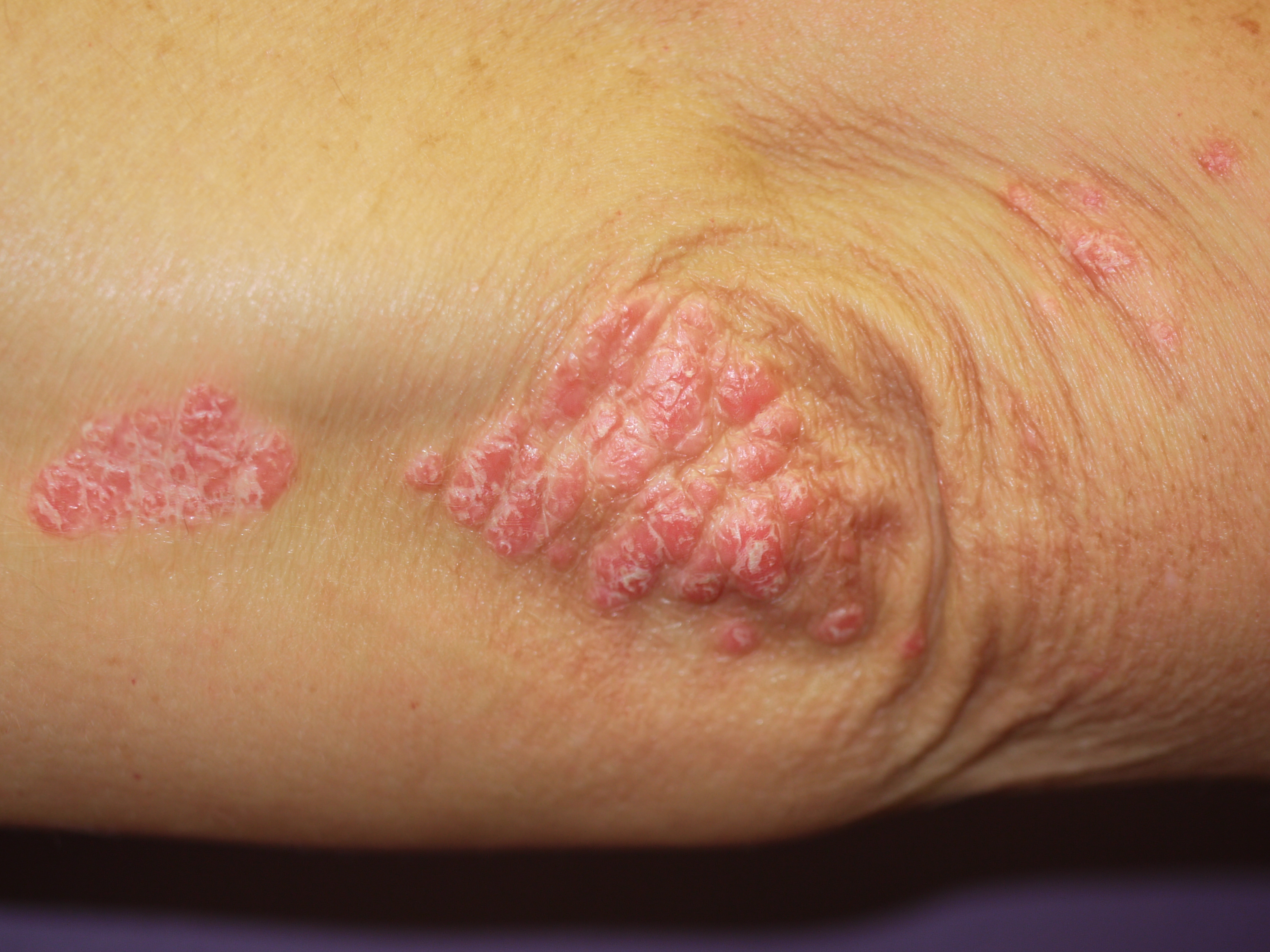
Actinic keratoses, also known as solar keratoses, are dry scaly patches of skin caused by damage from years of sun exposure.
The patches can be pink, red or brown in colour, and can vary in size from a few millimetres to a few centimetres across. The skin in affected areas can sometimes become very thick, and occasionally the patches can look like small horns or spikes.
Actinic keratoses are found on areas of skin that are exposed to the sun, such as the:
face, especially the nose and forehead
forearms and backs of hands
in men, on the rims of the ears and bald scalps
in women, on the legs below the knees
The patches are usually harmless and sometimes get better on their own, but they can be sore, itchy and look unsightly. There is also a small risk that the patches could develop into a type of skin cancer called squamous cell carcinoma if they’re not treated.
You should see your GP if you think you may have actinic keratoses, so they can discuss treatment options with you.
Who is affected
Actinic keratoses are most commonly seen in fair-skinned people, especially those with blue eyes, red hair, freckles and a tendency to burn easily in the sun. Men are affected more often than women.
People who have lived or worked abroad in a sunny place, or who have worked outdoors or enjoy outdoor hobbies, are most at risk.
It may take many years before actinic keratoses develop – they don’t usually appear before the age of 40.
Studies carried out in the UK have suggested that around one in every four or five people over the age of 60 has actinic keratoses.
Diagnosing actinic keratoses
Your GP may be able to diagnose actinic keratoses by examining the patches on your skin.
In some cases, the diagnosis may need to be confirmed by removing a small sample of skin and examining it under the microscope.
Actinic keratoses can often be managed by your GP, but you may need to see a skin specialist (dermatologist) for further assessment if:
your GP is not certain about your diagnosis
your GP thinks one or more of your patches may be cancerous or at a high risk of becoming cancerous
your patches are particularly severe or widespread
you are taking immunosuppressant drugs – for example, following an organ transplant
your patches have not responded to treatment
Treatment options
If the patches are not troublesome, your doctor may simply recommend that you keep an eye on them and come back if they change in any way – for example, if you develop new symptoms such as a patch growing quickly, bleeding or forming an ulcer.
However, actinic keratoses are often removed because of concerns they may develop into skin cancer (see below) or, less commonly, for cosmetic reasons.
The patches can be removed using a variety of treatments, depending on your individual circumstances. The main treatments used are summarised below.
Creams or gels
There are a number of creams and gels that can be applied to the skin if you have several patches. Commonly used treatments include 5-fluorouracil cream, imiquimod cream, diclofenac gel and ingenol mebutate gel.
These creams and gels are usually applied daily (washing your hands carefully after), often for several weeks, and they cause the abnormal skin cells to die. They may make the skin sore, and it may weep and blister after a few days of treatment.
The various creams and gels seem to be similarly effective in treating actinic keratoses, although the potential side effects and the length of time that treatment is needed differs between each of them. Not all are easily available.
Discuss the benefits and risks of the different creams and gels available with your GP before starting treatment.
Freezing with liquid nitrogen (cryotherapy)
In some cases, freezing the patches (cryotherapy) may be recommended. This causes blistering and shedding of the sun-damaged areas of skin.
The time it takes the skin to heal varies, depending on the areas of the body treated. Some areas may heal in a week or two, whereas others may take a few months to fully heal.
A light freeze usually leaves no scar, but thicker lesions or early skin cancer may need longer freezes, which can leave a permanently pale or dark mark.
Scraping (curettage)
Curettage is where the abnormal patches are scraped off with a sharp spoon-like instrument called a curette. This procedure is done under a local anaesthetic (where the treated area is numbed) and is generally used to treat thicker patches and early skin cancers, or to help confirm a diagnosis.
Cautery (heat treatment) is used to stop any bleeding after the cells have been removed. A scab forms after the procedure, which heals over a few weeks to leave a small scar.
The scrapings that are removed can be examined under the microscope to confirm the diagnosis.
Cutting it out (excision)
If your doctor suspects the patch may be cancerous or pre-cancerous, they may cut it out using a scalpel under local anaesthetic and close the wound with stitches. The piece of skin is then examined under the microscope to confirm the diagnosis.
Removing the patch will leave a permanent scar.
Other treatments
There are also a number of other treatments that may be effective in treating actinic keratoses, including:
photodynamic therapy (PDT) – where light is shone onto the affected area of skin after a light-activated cream has been applied; the light activates this cream and causes it to form a chemical that kills the abnormal cells
laser resurfacing – where a laser beam is used to remove the abnormal patches of skin
dermabrasion – where specially-designed abrasive instruments are used to remove the abnormal patches
chemical peels – where a corrosive liquid is applied to the affected area of skin to remove the abnormal patches
However, these treatments are not in widespread use and there is no clear evidence that they offer any additional benefit.
Self-help
It is important to protect your skin from the sun if you have actinic keratoses. This can reduce the risk of further patches developing and may help reduce the number of patches you already have.
To protect yourself from the sun, you should:
apply sunscreen with a sun protection factor (SPF) of at least 15 before exposing yourself to direct sunlight
cover up your skin with clothes and a hat during the summer months
try to avoid direct exposure to the sun when it is at its strongest (between 11am and 3pm)
It may also be helpful to regularly use emollients on your skin to stop it becoming dry.
Outlook
Actinic keratoses that have been treated usually go away, but it is likely that more patches will develop, requiring further treatment.
The development of actinic keratoses is a sign that the underlying skin is damaged from many years of sun exposure, and this cannot be reversed. It means you have a higher than average risk of developing skin cancer.
However, the exact chances of actinic keratoses developing into skin cancer are not clear. Some research has suggested the chances of a patch becoming cancerous are less than 1 in 1,000 every year, whereas other studies suggest the overall chances of actinic keratoses becoming cancerous may be as high as 1 in 10.


