Autism may develop in the womb
Children with autism may have too many cells in the brain regions responsible for emotional development, the Daily Mail has reported. The newspaper also said that, so far, genetics appears to be involved in less than a fifth of cases. It suggests that new research is pointing towards environmental factors, possibly in the womb, as a potential cause of the condition.
The intriguing research behind this news will no doubt be of interest to both scientists and parents of children with autism. However, the study itself was small, looking at post-mortem brain tissue taken from just seven boys with autism and six boys without the condition. The research found that in this small pool of samples children with autism had 67% more neurones (brain cells) within regions that deal with emotion and decision-making. They also found that the brains from the children with autism had a greater brain weight for age than would be expected.
This study should be considered to be preliminary, and will need following up to see if the phenomenon is present in further tissue samples. If it is found to be common among children with autism then the next steps will be to determine how it affects the workings of the brain and what actually causes it to occur.
Where did the story come from?
The study was carried out by researchers from the University of California, San Diego and other US universities. It was funded by several charitable organisations and research groups, including Autism Speaks, Cure Autism Now, the Peter Emch Family Foundation, the Simons Foundation, The Thursday Club Juniors and the University of California.
The study was published in the peer-reviewed Journal of the American Medical Association (JAMA).
The study was covered appropriately by the Daily Mail, but it is still not clear how much genetic or environmental causes have contributed to the differences that the researchers found. The Independent gave a brief but appropriate summary of this research.
What kind of research was this?
This research compared the anatomy of post-mortem brain samples from male children with and without autism in order to detect if there were any structural differences.
The researchers were looking for evidence of “brain overgrowth”, a phenomenon where children with autism possess certain regions of the brain that are larger than average. The researchers say that some studies have observed brain overgrowth in children with autism even before clinical signs appear, and particularly in an area at the front of the brain called the prefrontal cortex. The prefrontal cortex is thought to play a role in complicated behaviours such as the expression of personality, decision-making and governing appropriate social behaviour.
The researchers say that the anatomical structure of the brain overgrowth is currently unclear and so wanted to look at what type of brain cells were present in these areas. Types of brain cells include neurones, which pass messages between each other, and “glial” cells, which provide support functions to neurones.
What did the research involve?
The researchers obtained post-mortem brains from various university tissue banks where people had donated their children’s brain tissue for subsequent research.
They obtained brain samples from seven male children with autism and six without autism (the control group), all aged from 2 to 16 years old who had had their brains donated to science. As post-mortem tissue from younger individuals is rare, the researchers examined all control samples available to them at the time and nearly all of the autism samples available in their tissue banks. Most of the children had died in accidents where their brains had been starved of oxygen, for example by drowning.
The researchers recorded what the cause of death had been, how long the sample had been in storage and the individual’s ethnicity. They also interviewed their next of kin using a recognised diagnostic interview for autism, to determine what type of autism the child had.
The researchers then counted the number of neurone-type brain cells in the front regions of the brain samples. They also weighed the brains and compared their weights to age-expected norms (derived using data from 11,000 cases across 10 other brain weight studies designed to determine average weights for each age).
What were the basic results?
Through the next-of-kin interviews, all the children with autism were confirmed as definitely having had a full autistic disorder, according to reliable scales. None of the children had Asperger’s syndrome, which is typically a milder condition within the autistic spectrum. One seven-year-old in the autism group had a history of seizures requiring medication, and one seven-year-old in the control group had been taking medication for hyperactivity.
Compared with brain-weight norms, the brain weights of the children with autism were 17.6% heavier than average (95% CI, 10.2% to 25.0%; p=0.001). The brain weights of the control cases were not heavier than average for their respective ages.
The children with autism had 67% more neurones in the prefrontal cortex compared with the control children: 1.94 billion cells on average, compared with an average 1.16 billion in control subjects (95% CI 1.57 to 2.31 versus 95% CI 0.90 to 1.42).
How did the researchers interpret the results?
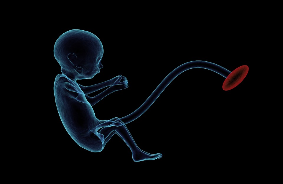
The researchers say that their preliminary study showed that children with autism may have a greater number of neurones in key frontal regions of their brains. They say that new neurones are not generated after birth, which means that this increased number of neurones must have come about before birth. They suggest that during development in the womb the excess number could have occurred because of more neurones developing unchecked, or through fewer neurones dying during this time.
Conclusion
This small, preliminary study looked at the anatomical features in brains of children who had autism and compared them to post-mortem brains from children without autism. In the small range of samples tested the researchers found that children with autism had around two-thirds more neurone brain cells in the front region of their brain than children without autism. They also found that when they compared the weight of their brains with age-adjusted norms, the children with autism had heavier brain weights than expected.
These results will no doubt be of great interest to both researchers and the parents of children with autism. However, one major limitation of this study must be taken into account: the availability of brain samples for research from children who have died is, understandably, low. This means that this research could only compare seven children who had autism with six children without autism. Having so few samples to compare means we cannot be certain if this type of brain overgrowth is typical of autistic children or simply due to chance findings.
Beyond this limitation, the researchers have described the characteristics of these children but it is possible that children with autism who die from accidents may in some way differ from other children with autistic spectrum disorder which made them more likely to suffer accidents. It is not clear whether the same pattern of overgrowth would be observed in a larger sample and so care should be taken in assuming that these results apply to all children with autistic spectrum disorders.
The researchers have suggested that new neurones in this area of the brain are not generated after birth, and that the increased number of cells in autistic brains suggests that either there is above-average production of these cells while the children were in the womb, or less-than-average programmed death of these cells after birth to regulate cell numbers. Even though we are born with a set number of neurones, the neurones can continue to form new branches that join them with other neurones. The number and strength of these connections between neurones is important in determining how our brain functions.
In short, this study only looked at a small number of samples and should be considered to be preliminary. Its intriguing results will now need to be followed up to see if the effects are seen in further samples and also to tell exactly why the phenomenon might occur. For example, we cannot yet tell whether genetic or environmental mechanisms are behind the relationship or exactly how these changes in brain structure might cause the behaviours seen in people with autism.


