When muscles work out, they help neurons to grow, a new study shows

Exercising Neurons

MIT scientists discovered that motor neuron growth significantly increased over five days in response to exercise-related biochemical (left) and mechanical (right) signals. The green ball represents a neuron cluster that extends outward with long tails, known as axons. Credit: Angel Bu.

Exercise is undoubtedly beneficial for the body. Regular physical activity strengthens muscles and enhances bones, blood vessels, and the immune system.

Recent research by MIT engineers has revealed that exercise can also benefit individual neurons. They discovered that when muscles contract during physical activity, they release biochemical signals known as myokines. Neurons exposed to these muscle-generated signals grew up to four times farther than those not exposed to myokines. This cellular-level research indicates that exercise can have a significant biochemical impact on nerve growth.

Researchers made an intriguing discovery: neurons respond not only to the biochemical signals released during exercise but also to the physical stress that occurs during it. They found that when neurons are stretched and then released repeatedly—similar to how muscles contract and expand during physical activity—these neurons grow significantly, just as they do when exposed to myokines produced by the muscles.

Previous studies have suggested a possible biochemical link between muscle activity and nerve growth. However, according to the researchers, this study is the first to demonstrate that physical effects can be equally significant. The findings, set to be published in the journal *Advanced Healthcare Materials*, highlight the relationship between muscles and nerves during exercise. This could lead to the development of exercise-related therapies to repair damaged and deteriorating nerves.

“Now that we understand the existence of muscle-nerve crosstalk, this knowledge could be beneficial for treating conditions such as nerve injuries, where communication between nerves and muscles is disrupted,” explains Ritu Raman, the Eugene Bell Career Development Assistant Professor of Mechanical Engineering at MIT. “By stimulating the muscle, we may be able to encourage the nerve to heal, potentially restoring mobility to individuals who have lost it due to traumatic injuries or neurodegenerative diseases.”

Muscle talk

In 2023, Raman and her colleagues reported that they could restore mobility in mice that had experienced a traumatic muscle injury by implanting muscle tissue at the injury site and then exercising the new tissue by stimulating it repeatedly with light. Over time, they found that the exercised graft helped mice regain their motor function, reaching activity levels comparable to those of healthy mice. 

When the researchers analyzed the graft itself, regular exercise appeared to stimulate the grafted muscle to produce specific biochemical signals that promote nerve and blood vessel growth. 

“That was interesting because we always think that nerves control muscle, but we don’t think of muscles talking back to nerves,” Raman says. “So, we started to think stimulating muscle was encouraging nerve growth. People replied that maybe that’s the case, but there are hundreds of other cell types in an animal, and it’s hard to prove that the nerve is growing more because of the muscle rather than the immune system or something else playing a role.”

In their new study, the team focused solely on muscle and nerve tissue to determine whether exercising muscles directly affects nerve growth. The researchers grew mouse muscle cells into long fibres that then fused to form a small sheet of mature muscle tissue about the size of a quarter. 

The team genetically modified the muscle to contract in response to light. With this modification, the team could flash a light repeatedly, causing the muscle to squeeze in response, mimicking the act of exercise. Raman previously developed a novel gel mat to grow and exercise muscle tissue. The gel’s properties are such that it can support muscle tissue and prevent it from peeling away as the researchers stimulated the muscle to exercise. 

The team then collected samples of the surrounding solution in which the muscle tissue was exercised, thinking that the solution should hold myokines, including growth factors, RNA, and a mix of other proteins. 

“I would think of myokines as a biochemical soup of things that muscles secrete, some of which could be good for nerves and others that might have nothing to do with nerves,” Raman says. “Muscles are pretty much always secreting myokines, but when you exercise them, they make more.”

“Exercise as medicine”

The team transferred the myokine solution to a separate dish containing motor neurons — nerves found in the spinal cord that control muscles involved in voluntary movement. The researchers grew the neurons from stem cells derived from mice. As with the muscle tissue, the neurons were grown on a similar gel mat. After the neurons were exposed to the myokine mixture, the team observed that they quickly began to grow, four times faster than neurons that did not receive the biochemical solution. 

“They grow much farther and faster, and the effect is pretty immediate,” Raman notes. 

To examine how neurons changed in response to exercise-induced myokines more closely, the team performed a genetic analysis, extracting RNA from the neurons to determine whether the myokines induced any change in the expression of certain neuronal genes. 

“We saw that many of the genes up-regulated in the exercise-stimulated neurons was not only related to neuron growth, but also neuron maturation, how well they talk to muscles and other nerves, and how mature the axons are,” Raman says. “Exercise seems to impact not just neuron growth but also how mature and well-functioning they are.” 

The results suggest that exercise’s biochemical effects can promote neuron growth. The group then wondered If exercise’s purely physical impacts could have a similar benefit. 

“Neurons are physically attached to muscles, so they are also stretching and moving with the muscle,” Raman says. “We also wanted to see, even in the absence of biochemical cues from muscle, could we stretch the neurons back and forth, mimicking the mechanical forces (of exercise), and could that have an impact on growth as well?”

To answer this, the researchers grew a different set of motor neurons on a gel mat that they embedded with tiny magnets. They then used an external magnet to jiggle the mat — and the neurons — back and forth. In this way, they “exercised” the neurons, for 30 minutes a day. To their surprise, they found that this mechanical exercise stimulated the neurons to grow just as much as the myokine-induced neurons, growing significantly farther than neurons that received no form of exercise. 

“That’s a good sign because it tells us both biochemical and physical effects of exercise are equally important,” Raman says. 

Now that the group has shown that exercising muscle can promote nerve growth at the cellular level, they plan to study how targeted muscle stimulation can be used to grow and heal damaged nerves and restore mobility for people with neurodegenerative diseases such as ALS.

“This is just our first step toward understanding and controlling exercise as medicine,” Raman says. 

Drug reverses age-related cognitive decline within days


Rapid mental rejuvenation in old mice suggests age-related losses may be broadly reversible 

"Limiting children's screen time linked to better cognition," reports BBC News.


Just a few doses of an experimental drug can reverse age-related declines in memory and mental flexibility in mice, according to a new study by UC San Francisco scientists. The drug, called ISRIB, has already been shown in laboratory studies to restore memory function months after traumatic brain injury (TBI), reverse cognitive impairments in Down Syndrome , prevent noise-related hearing loss, fight certain types of prostate cancer , and even enhance cognition in healthy animals.

In the new study, published December 1, 2020 in the open-access journal eLife , researchers showed rapid restoration of youthful cognitive abilities in aged mice, accompanied by a rejuvenation of brain and immune cells that could help explain improvements in brain function.


“ISRIB’s extremely rapid effects show for the first time that a significant component of age-related cognitive losses may be caused by a kind of reversible physiological “blockage” rather than more permanent degradation,” said Susanna Rosi , PhD, Lewis and Ruth Cozen Chair II and professor in the departments of Neurological Surgery and of Physical Therapy and Rehabilitation Science (http://ptrehab.ucsf.edu/) .

“The data suggest that the aged brain has not permanently lost essential cognitive capacities, as was commonly assumed, but rather that these cognitive resources are still there but have been somehow blocked, trapped by a vicious cycle of cellular stress,” added Peter Walter , PhD, a professor in the UCSF Department of Biochemistry and Biophysics and a Howard Hughes Medical Institute investigator. “Our work with ISRIB demonstrates a way to break that cycle and restore cognitive abilities that had become walled off over time.”


Could Rebooting Cellular Protein Production Hold the Key to Aging and Other Diseases?

Walter has won numerous scientific awards, including the Breakthrough , Lasker  and Shaw  prizes, for his decades-long studies of cellular stress responses. ISRIB, discovered in 2013 in Walter’s lab, works by rebooting cells’ protein production machinery after it gets throttled by one of these stress responses — a cellular quality control mechanism called the integrated stress response (ISR; ISRIB stands for ISR InhiBitor).

The ISR normally detects problems with protein production in a cell — a potential sign of viral infection or cancer-promoting gene mutations — and responds by putting the brakes on cell’s protein-synthesis machinery. This safety mechanism is critical for weeding out misbehaving cells, but if stuck in the on position in a tissue like the brain, it can lead to serious problems, as cells lose the ability to perform their normal activities, Walter and colleagues have found.

In particular, recent animal studies by Walter and Rosi, made possible by early philanthropic support from The Rogers Family Foundation, have implicated chronic ISR activation in the persistent cognitive and behavioral deficits seen in patients after TBI, by showing that, in mice, brief ISRIB treatment can reboot the ISR and restore normal brain function almost overnight.

The cognitive deficits in TBI patients are often likened to premature aging, which led Rosi and Walter to wonder if the ISR could also underlie purely age-related cognitive decline. Aging is well known to compromise cellular protein production across the body, as life’s many insults pile up and stressors like chronic inflammation wear away at cells, potentially leading to widespread activation of the ISR.

“We’ve seen how ISRIB restores cognition in animals with traumatic brain injury, which in many ways is like a sped-up version of age-related cognitive decline,” said Rosi, who is director of neurocognitive research in the UCSF Brain and Spinal Injury Center and a member of the UCSF Weill Institute for Neurosciences. “It may seem like a crazy idea, but asking whether the drug could reverse symptoms of aging itself was just a logical next step.”

ISRIB Improves Cognition, Boosts Neuron and Immune Cell Function

In the new study, researchers led by Rosi lab postdoc Karen Krukowski , PhD, trained aged animals to escape from a watery maze by finding a hidden platform, a task that is typically hard for older animals to learn. But animals who received small daily doses of ISRIB during the three-day training process were able to accomplish the task as well as youthful mice, much better than animals of the same age who didn’t receive the drug.

The researchers then tested how long this cognitive rejuvenation lasted and whether it could generalize to other cognitive skills. Several weeks after the initial ISRIB treatment, they trained the same mice to find their way out of a maze whose exit changed daily — a test of mental flexibility for aged mice who, like humans, tend to get increasingly stuck in their ways. The mice who had received brief ISRIB treatment three weeks before still performed at youthful levels, while untreated mice continued to struggle.

To understand how ISRIB might be improving brain function, the researchers studied the activity and anatomy of cells in the hippocampus, a brain region with a key role in learning and memory, just one day after giving animals a single dose of ISRIB. They found that common signatures of neuronal aging disappeared literally overnight: neurons’ electrical activity became more sprightly and responsive to stimulation, and cells showed more robust connectivity with cells around them while also showing an ability to form stable connections with one another usually only seen in younger mice.

The researchers are continuing to study exactly how the ISR disrupts cognition in aging and other conditions and to understand how long ISRIB’s cognitive benefits may last. Among other puzzles raised by the new findings is the discovery that ISRIB also alters the function of the immune system’s T cells, which also are prone to age-related dysfunction. The findings suggest another path by which the drug could be improving cognition in aged animals, and could have implications for diseases from Alzheimer’s to diabetes that have been linked to heightened inflammation caused by an aging immune system.

“This was very exciting to me because we know that aging has a profound and persistent effect on T cells and that these changes can affect brain function in the hippocampus,” said Rosi. “At the moment, this is just an interesting observation, but it gives us a very exciting set of biological puzzles to solve.

ISRIB May Have Wide-Ranging Implications for Neurological Disease

It turns out that chronic ISR activation and resulting blockage of cellular protein production may play a role in a surprisingly wide array of neurological conditions. Below is a partial list of these conditions, based on a recent review by Walter and colleague Mauro Costa-Mattioli of Baylor College of Medicine, which could potentially be treated with an ISR-resetting agent like ISRIB: 

  • Frontotemporal Dementia
  • Alzheimer’s Disease
  • Amyotrophic Lateral Sclerosis (ALS)
  • Age-related Cognitive Decline
  • Multiple Sclerosis
  • Traumatic Brain Injury
  • Parkinson’s Disease
  • Down Syndrome
  • Vanishing White Matter Disorder
  • Prion Disease

ISRIB has been licensed by Calico, a South San Francisco, Calif. company exploring the biology of aging, and the idea of targeting the ISR to treat disease has been picked up by other pharmaceutical companies, Walter says.

One might think that interfering with the ISR, a critical cellular safety mechanism, would be sure to have serious side effects, but so far in all their studies, the researchers have observed none. This is likely due to two factors, Walter says. First, it takes just a few doses of ISRIB to reset unhealthy, chronic ISR activation back to a healthier state, after which it can still respond normally to problems in individual cells. Second, ISRIB has virtually no effect when applied to cells actively employing the ISR in its most powerful form — against an aggressive viral infection, for example.

Naturally, both of these factors make the molecule much less likely to have negative side effects — and more attractive as a potential therapeutic. According to Walter: “It almost seems too good to be true, but with ISRIB we seem to have hit a sweet spot for manipulating the ISR with an ideal therapeutic window.


Have you taken the Ice Bucket Challenge yet? What do you think of it as a way of raising awareness of ALS (Motor Neurone Disease)?


President Bush and the Ice Bucket Challenge

President Bush and the Ice Bucket Challenge

The Ice Bucket challenge had gone far more than viral by the time I picked up on it. I was away on holiday so was not really looking at social media.

But by the time we got home it was in full swing. And as with all good thing it seemed to have become very controversial.

Now as some readers will know amyotrophic lateral sclerosis (ALS), which is the most common form of Motor Neurone Disease was the medical condition from which my mother-in-law passed way some four years ago! If you wish to make a donation to her local ALS charity you can do so here.

What interests me is how many of us have done the ice bucket challenge so far. Please share in the poll below. If you could tell us which cause you donated to and a link in the comments box that would be great!


Many thanks in advance.


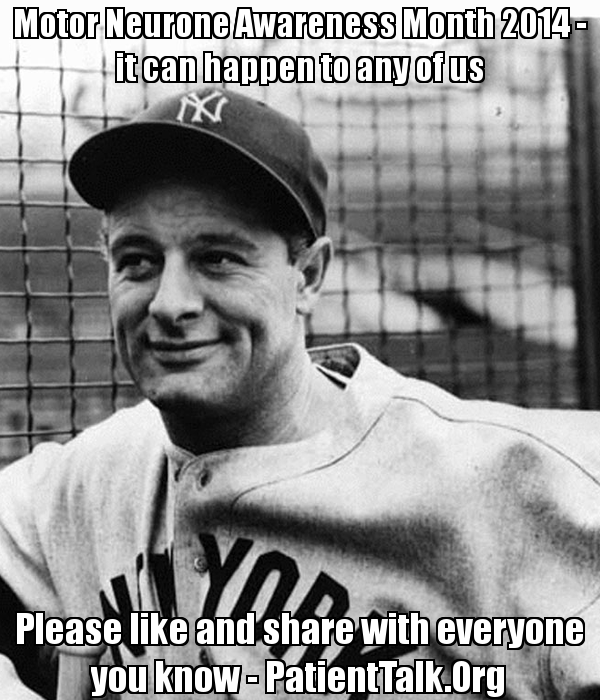
Motor Neurone Disease Awareness Month 2014 – please like and share to help raise awareness of MND, ALS or Lou Gehrig’s Disease #MND


Motor Neurone Awareness Month 2014 - Lou Gehrig

Motor Neurone Awareness Month 2014 – Lou Gehrig

This man was Lou Gehrig. He gave his name to the most common type of Motor Neurone Disease called Amyotrophic lateral sclerosis or ALS.

While , of course, famous people so get MND, so do the rest of us. This post is dedicated to the memory of my late mother in law who died this month five years ago after battling ALS for three years.

A few months ago on old friend told me that his mother had just been diagnosed with MND. So this is a cause very dear to my heart.

So far there is no cure just therapies for easing the effects on the condition. So for many people with MND and their loved ones the situation can feel very hopeless.

I would be very grateful if you could share this page and image to help raise awareness of Motor Neurone Disease.

For more information on the month itself please go to the web site of the Motor Neurone Disease Association.

Many thanks in advance and just to let you know that the hashtag for the month is #MND