In our morning rounds, a new approach to fight multiple sclerosis — a disease where the body attacks its own central nervous system. MS affects about 400,000 people in the United States. It is two to three times more common among women. Current treatments may have severe side effects, and there is no cure. Dr. Tara Narula joins “CBS This Morning” to discuss a cutting edge, but low-tech attempt to slow the symptoms.
When Dr Chris van Tulleken embarked on an ultra-processed 30 day diet to uncover what effect it has on our bodies, the results leave him and the scientists in shock!
Neuroscientists link autism to reduced activity of key neurotransmitter in human brain
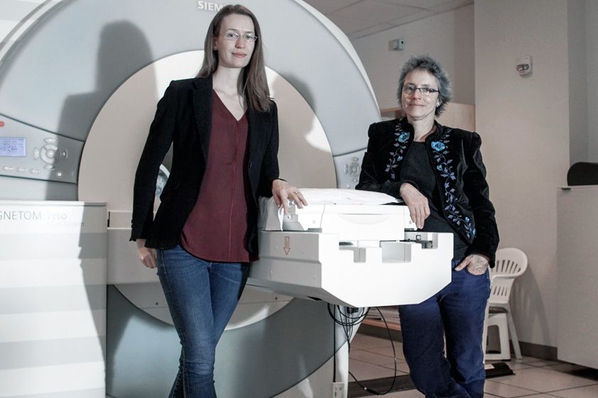
Caption:(Left to right) Caroline Robertson and Nancy Kanwisher.Credits:Photo: Sham Sthankiya
MIT and Harvard University neuroscientists have found a link between a behavioral symptom of autism and reduced activity of a neurotransmitter whose job is to dampen neuron excitation. The findings suggest that drugs that boost the action of this neurotransmitter, known as GABA, may improve some of the symptoms of autism, the researchers say.
Brain activity is controlled by a constant interplay of inhibition and excitation, which is mediated by different neurotransmitters. GABA is one of the most important inhibitory neurotransmitters, and studies of animals with autism-like symptoms have found reduced GABA activity in the brain. However, until now, there has been no direct evidence for such a link in humans.
“This is the first connection in humans between a neurotransmitter in the brain and an autistic behavioral symptom,” says Caroline Robertson, a postdoc at MIT’s McGovern Institute for Brain Research and a junior fellow of the Harvard Society of Fellows. “It’s possible that increasing GABA would help to ameliorate some of the symptoms of autism, but more work needs to be done.”
Robertson is the lead author of the study, which appears in the Dec. 17 online edition of Current Biology. The paper’s senior author is Nancy Kanwisher, the Walter A. Rosenblith Professor of Brain and Cognitive Sciences and a member of the McGovern Institute. Eva-Maria Ratai, an assistant professor of radiology at Massachusetts General Hospital, also contributed to the research.
Too little inhibition
Many symptoms of autism arise from hypersensitivity to sensory input. For example, children with autism are often very sensitive to things that wouldn’t bother other children as much, such as someone talking elsewhere in the room, or a scratchy sweater. Scientists have speculated that reduced brain inhibition might underlie this hypersensitivity by making it harder to tune out distracting sensations.
In this study, the researchers explored a visual task known as binocular rivalry, which requires brain inhibition and has been shown to be more difficult for people with autism. During the task, researchers show each participant two different images, one to each eye. To see the images, the brain must switch back and forth between input from the right and left eyes.
For the participant, it looks as though the two images are fading in and out, as input from each eye takes its turn inhibiting the input coming in from the other eye.
“Everybody has a different rate at which the brain naturally oscillates between these two images, and that rate is thought to map onto the strength of the inhibitory circuitry between these two populations of cells,” Robertson says.
She found that nonautistic adults switched back and forth between the images nine times per minute, on average, and one of the images fully suppressed the other about 70 percent of the time. However, autistic adults switched back and forth only half as often as nonautistic subjects, and one of the images fully suppressed the other only about 50 percent of the time.
Performance on this task was also linked to patients’ scores on a clinical evaluation of communication and social interaction used to diagnose autism: Worse symptoms correlated with weaker inhibition during the visual task.
The researchers then measured GABA activity using a technique known as magnetic resonance spectroscopy, as autistic and typical subjects performed the binocular rivalry task. In nonautistic participants, higher levels of GABA correlated with a better ability to suppress the nondominant image. But in autistic subjects, there was no relationship between performance and GABA levels. This suggests that GABA is present in the brain but is not performing its usual function in autistic individuals, Robertson says.
“GABA is not reduced in the autistic brain, but the action of this inhibitory pathway is reduced,” she says. “The next step is figuring out which part of the pathway is disrupted.”
“This is a really great piece of work,” says Richard Edden, an associate professor of radiology at the Johns Hopkins University School of Medicine. “The role of inhibitory dysfunction in autism is strongly debated, with different camps arguing for elevated and reduced inhibition. This kind of study, which seeks to relate measures of inhibition directly to quantitative measures of function, is what we really to need to tease things out.”
Early diagnosis
In addition to offering a possible new drug target, the new finding may also help researchers develop better diagnostic tools for autism, which is now diagnosed by evaluating children’s social interactions. To that end, Robertson is investigating the possibility of using EEG scans to measure brain responses during the binocular rivalry task.
“If autism does trace back on some level to circuitry differences that affect the visual cortex, you can measure those things in a kid who’s even nonverbal, as long as he can see,” she says. “We’d like it to move toward being useful for early diagnostic screenings.”
Loss of Shank gene prevents neuronal synapses from properly maturing
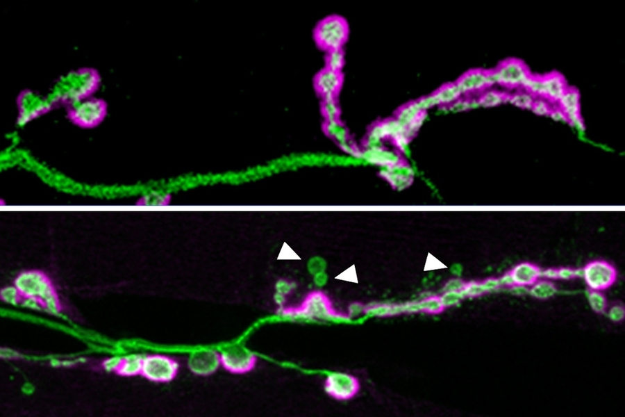
Caption:In the top image, a neuronal axon (green) forms many synapses, highlighted in purple. The bottom image shows an axon from a fruit fly lacking the Shank protein. In these flies, not as many synapses form, and some of them (indicated by the white arrows) do not fully mature.Credits:Image courtesy of the researchers.
A new study from MIT neuroscientists reveals that a gene mutation associated with autism plays a critical role in the formation and maturation of synapses — the connections that allow neurons to communicate with each other.
Many genetic variants have been linked to autism, but only a handful are potent enough to induce the disorder on their own. Among these variants, mutations in a gene called Shank3 are among the most common, occurring in about 0.5 percent of people with autism.
Scientists know that Shank3 helps cells respond to input from other neurons, but because there are two other Shank proteins, and all three can fill in for each other in certain ways, it has been difficult to determine exactly what the Shank proteins are doing.
“It’s clearly regulating something in the neuron that’s receiving a synaptic signal, but some people find one role and some people find another,” says Troy Littleton, a professor in the departments of Biology and of Brain and Cognitive Sciences at MIT, a member of MIT’s Picower Institute for Learning and Memory, and the senior author of the study. “There’s a lot of debate over what it really does at synapses.”
Key to the study is that fruit flies, which Littleton’s lab uses to study synapses, have only one version of the Shank gene. By knocking out that gene, the researchers eliminated all Shank protein from the flies.
“This is the first animal where we have completely removed all Shank family proteins,” says Kathryn Harris, a Picower Institute research scientist and lead author of the paper, which appears in the May 25 issue of the Journal of Neuroscience.
Synaptic organization
Scientists already knew that the Shank proteins are scaffold proteins, meaning that they help to organize the hundreds of other proteins found in the synapse of a postsynaptic cell — a cell that receives signals from a presynaptic cell. These proteins help to coordinate the cell’s response to the incoming signal.
“Shank is essentially a hub for signaling,” Harris says. “It brings in a lot of other partners and plays an organizational role at the postsynaptic membrane.”
In fruit flies lacking the Shank protein, the researchers found two dramatic effects. First, the postsynaptic cells had many fewer boutons, which are the sites where neurotransmitter release occurs. Second, many of the boutons that did form were not properly developed; that is, they were not surrounded by all of the postsynaptic proteins normally found there, which are required to respond to synaptic signals.
The researchers are now studying how this reduction in functional synapses affects the brain. Littleton suspects that the development of neural circuits could be impaired, which, if the same holds true in humans, may help explain some of the symptoms seen in autistic people.
“During critical windows of social and language learning, we reshape our connections to drive connectivity patterns that respond to rewards and language and social interactions,” he says. “If Shank is doing similar things in the mammalian brain, one could imagine potentially having those circuits form relatively normally early on, but if they fail to properly mature and form the proper number of connections, that could lead to a variety of behavioral defects.”
Pinpointing an exact link to autism symptoms would be difficult to do in fruit fly studies, however.
“Although the core molecular machines that allow neurons to communicate are highly conserved between fruit flies and humans, the anatomy of the various circuits that are formed during evolution are quite different,” Littleton says. “It’s hard to jump from a synaptic defect in the fly to an autism-like phenotype because the circuits are so different.”
An unexpected link
The researchers also showed, for the first time, that loss of Shank affects a well-known set of proteins that comprise the Wnt (also known as Wingless) signaling pathway. When a Wnt protein binds to a receptor on the cell, it initiates a series of interactions that influence which genes are turned on. This, in turn, contributes to many cell processes including embryonic development, tissue regeneration, and tumor formation.
When Shank is missing from fruit flies, Wnt signaling is disrupted because the receptor that normally binds to Wnt fails to be internalized by the cell. Normally, a small segment of the activated receptor moves to the cell nucleus and influences the transcription of genes that promote maturation of synapses. Without Shank, Wnt signaling is impaired and the synapses do not fully mature.
“The Shank protein and the Wnt protein family are thought to be involved in autism independently, but the fact that this study discovered that Wnt and Shank are interacting brings the story into better focus,” says Bryan Stewart, a professor of cell and systems biology at the University of Toronto at Mississauga, who was not involved in the research. “Now we can look and see if those interactions between Wnt and Shank are potentially responsible for their role in autism.”
The finding raises the possibility of treating autism with drugs that promote Wnt signaling, if the same connection is found in humans.
“Because the link to Wnt signaling is new and hasn’t been picked up in mammalian studies, we really hope that that can inspire people to look for a connection to Wnt signaling in mammalian models, and maybe that can offer another avenue for how loss of Shank could be counteracted,” Harris says.
The research was funded by the National Institutes of Health and the Simons Center for the Social Brain at MIT.
We use cookies on our website to give you the most relevant experience by remembering your preferences and repeat visits. By clicking “Accept”, you consent to the use of ALL the cookies.
This website uses cookies to improve your experience while you navigate through the website. Out of these, the cookies that are categorized as necessary are stored on your browser as they are essential for the working of basic functionalities of the website. We also use third-party cookies that help us analyze and understand how you use this website. These cookies will be stored in your browser only with your consent. You also have the option to opt-out of these cookies. But opting out of some of these cookies may affect your browsing experience.
Necessary cookies are absolutely essential for the website to function properly. This category only includes cookies that ensures basic functionalities and security features of the website. These cookies do not store any personal information.
Any cookies that may not be particularly necessary for the website to function and is used specifically to collect user personal data via analytics, ads, other embedded contents are termed as non-necessary cookies. It is mandatory to procure user consent prior to running these cookies on your website.