Study reveals how CBD counters epileptic seizures – important for members of the autistic community

Microscope image


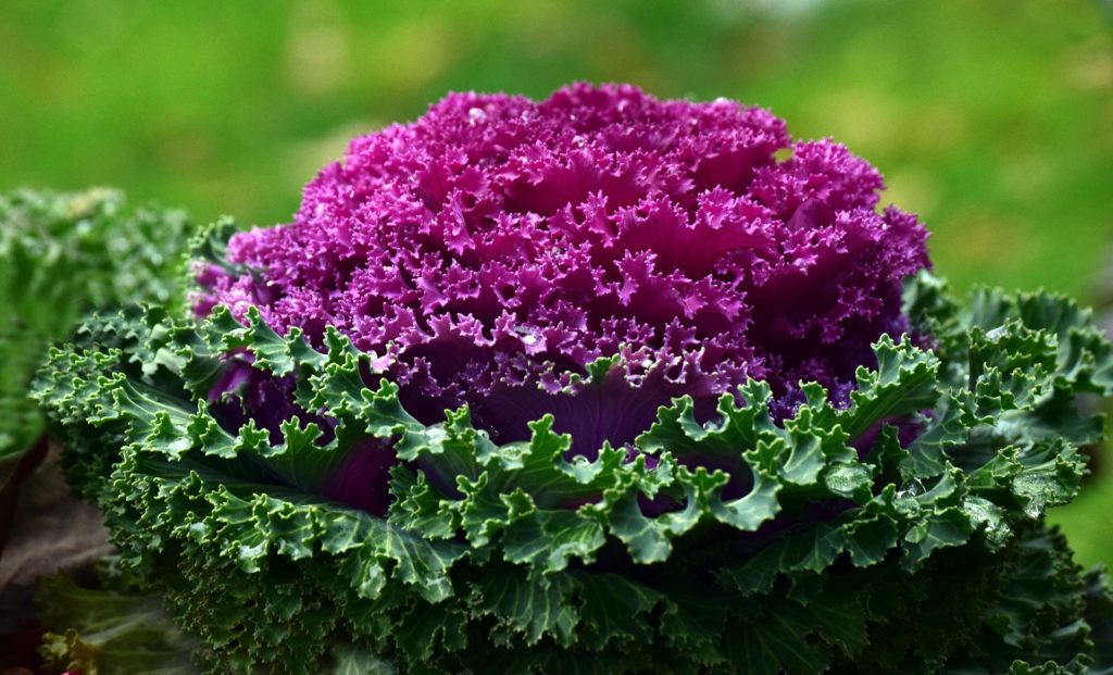
This microscope image of the brain region called the hippocampus shows the protein targeted by cannabis-derived CBD, GPR55 (red), and brain cells (blue) that send their extensions out to form the layers seen in the image. The interconnected nature of the hippocampus makes it a significant site for the initiation and spread of seizures.
Tsien et al, Courtesy of Cell Press

A study reveals a previously unknown way in which cannabidiol (CBD), a substance found in cannabis, reduces seizures in many treatment-resistant forms of pediatric epilepsy.  

Led by researchers at NYU Grossman School of Medicine, the new study found that CBD blocked signals carried by a molecule called lysophosphatidylinositol (LPI). Found in brain cells called neurons, LPI is thought to amplify nerve signals as part of normal function but can be hijacked by disease to promote seizures.

Published online February 13 in Neuron, the work confirmed a previous finding that CBD blocks the ability of LPI to amplify nerve signals in a brain region called the hippocampus. The current findings argue for the first time that LPI also weakens signals that counter seizures, further explaining the value of CBD treatment.   

“Our results deepen the field’s understanding of a central seizure-inducing mechanism, with many implications for the pursuit of new treatment approaches,” said corresponding author Richard W. Tsien, chair of the Department of Physiology and Neuroscience at NYU Langone Health.

“The study also clarified, not just how CBD counters seizures, but more broadly, how circuits are balanced in the brain,” added Tsien. “Related imbalances are present in autism and schizophrenia so that the paper may have a broader impact.”

Disease-Causing Loop

The study results build on how each neuron “fires” to send an electrical pulse down an extension of itself until it reaches a synapse, the gap that connects it to the next cell in a neuronal pathway. When it reaches the cell’s end before the synapse, the pulse triggers the release of compounds called neurotransmitters that float across the gap to affect the next cell in line. Upon crossing, such signals either encourage the cell to fire (excitation), or apply the brakes on firing (inhibition). Balance between the two are essential to brain function; too much excitation promotes seizures.   

The new study looked at several rodent models to explore mechanisms behind seizures, often by measuring information-carrying electrical current flows with fine-tipped electrodes. Other experiments looked at the effect of LPI by genetically removing its main signaling partner, or by measuring the release of LPI following seizures.

The tests confirmed past findings that LPI influences nerve signals by binding to a protein called G-coupled receptor 55 (GPR55), on neuron cell surfaces. This LPI-GPR55 presynaptic interaction was found to cause the release of calcium ions within the cell, which encouraged cells to release glutamate, the main excitatory neurotransmitter. Further, when LPI activated GPR55 on the other side of the synapse, it weakened inhibition, by decreasing the supply and proper arrangement of proteins necessary for inhibition. Collectively, this creates a “dangerous” two-pronged mechanism to increase excitability, say the authors.  

The research team found that either genetically engineering mice to lack GPR55, or treating mice with plant-derived CBD prior to seizure-inducing stimuli, blocked LPI-mediated effects on both excitatory and inhibitory synaptic transmission. While prior studies had implicated GPR55 as a seizure-reducing target of CBD, the current work provided a more detailed, proposed mechanism of action.

Purple vegetables and tubers have anti-diabetic properties

Purple vegetables and tubers have antidiabetic properties


The red, purple and blue pigments in fruits, vegetables, and tubers called anthocyanins can reduce the risk of diabetes by affecting energy metabolism, gut microbiota, and inflammation. A new review article comparing the research results shows that anthocyanins’ beneficial effect on type 2 diabetes is increased if the anthocyanin is acylated, meaning that an acyl group is added to the sugar moieties of anthocyanin.

A significant amount of acylated anthocyanins can be found in purple potatoes, purple sweet potatoes, radishes, purple carrots and red cabbages, whereas bilberries and mulberries contain mostly nonacylated anthocyanins. Acylated anthocyanins are poorly absorbed in digestion, but they have probiotic properties and reduce the risk of diabetes more efficiently than nonacylated anthocyanins.

“The studies have shown that, in addition to changing physical and chemical properties, the acylation affects how the anthocyanins are absorbed and metabolised,” says Postdoctoral Researcher Kang Chen at Food Sciences Unit, University of Turku, Finland.

The acylated anthocyanins are more effective antioxidants than the nonacylated anthocyanins, and they can also improve the intestinal barrier that enables the absorption of necessary nutrients. Furthermore, the acylated anthocyanins maintain gut microbiota homeostasis, suppress pro-inflammatory pathways, and modulate glucose and lipid metabolisms.

“The plant’s genotype defines what kind of anthocyanins they produce. In general, purple vegetables contain many acylated anthocyanins. Also, purple potatoes, especially the Finnish variety called ‘Synkeä Sakari’, is abundant in acylated anthocyanins,” says Chen.

Acylated anthocyanins travel through our bodies from the upper gastrointestinal tract to the colon where they are metabolised by the gut microbiota. Glucose transporters are involved in anthocyanin absorption, but different glucose transporters are responsible for the absorption of acylated and nonacylated anthocyanins. The acylated and nonacylated anthocyanins also have different impacts on the enzymes involved in metabolism.

“The latest research has shown that the acylated and nonacylated anthocyanins can impact type 2 diabetes in different ways,” Chen summarises.

Verified: COVID-19 infection increases diabetes risk – have you developed diabetes after COVID?


Investigators in the Smidt Heart Institute at Cedars-Sinai have confirmed that people who have had COVID-19 have an increased risk for new-onset diabetes—the most significant contributor to cardiovascular disease.

“Our results verify that the risk of developing Type 2 diabetes after a COVID-19 infection was not just an early observation but, in fact, a real risk that has, unfortunately, persisted through the Omicron era,” said Alan Kwan, MD, first and corresponding author of the study and a cardiovascular physician in the Smidt Heart Institute at Cedars-Sinai. 

The trend, Kwan says, is concerning because most people in the United States will eventually experience a COVID-19 infection. “This research study helps us understand—and better prepare for—the post-COVID-19 era of cardiovascular risk,” Kwan said. 

The findings, published today in the journal JAMA Network Open, also suggest that the risk of Type 2 diabetes appears lower in individuals who were already vaccinated against COVID-19 by the time they were infected. 

To determine the rising rates of diabetes, investigators evaluated medical records from 23,709 adult patients who had at least one documented COVID-19 infection and were treated within the Cedars-Sinai Health System in Los Angeles from 2020-2022. The average patient was 47 years old, and 54% of subjects were female.   

Within the study time frame:

  • The combined risk of Type 2 diabetes after COVID-19 exposure—accounting for both vaccinated and unvaccinated patients—was 2.1%, with 70% occurring after COVID-19 infection versus 30% happening prior to COVID-19 exposure.
  • The risk of Type 2 diabetes after COVID-19 exposure for unvaccinated patients was 2.7%, with 74% occurring after COVID-19 infection versus 26% happening prior to COVID-19 exposure.
  • The risk of Type 2 diabetes after COVID-19 exposure for vaccinated patients was 1.0%, with 51% occurring after COVID-19 infection versus 49% happening prior to COVID-19 exposure.

“These results suggest that COVID-19 vaccination prior to infection may provide a protective effect against diabetes risk,” said Kwan. “Although further studies are needed to validate this hypothesis, we remain steadfast in our belief that COVID-19 vaccination remains an important tool in protecting against COVID-19 and the still-uncertain risks that people may experience during the post-infection period.”   

Susan Cheng, MD, MPH, senior author of the study, professor of Cardiology, and director for Cardiovascular Population Sciences in the Smidt Heart Institute, says these findings broaden the medical field’s understanding of the effects of COVID-19 on the body, while simultaneously unearthing yet-to-be-answered questions. 

“Although we don’t yet know for certain, the trends and patterns that we see in the data suggest that COVID-19 infection could be acting in certain settings like a disease accelerator, amplifying risk for a diagnosis that individuals might have otherwise received later in life,” said Cheng, the Erika J. Glazer Chair in Women’s Cardiovascular Health and Population Science. “So, it could be that instead of being diagnosed with diabetes by age 65, a person with preexisting risk for diabetes might—after a COVID-19 infection—be more likely to develop diabetes by age 45 or 55.”

Diabetes disrupts normal metabolism and metabolic processes, preventing the pancreas from producing enough insulin, a hormone that helps regulate blood levels of glucose and amino acids. Because diabetes can damage vital organs and blood vessels, people with diabetes are at higher risk for heart attack and stroke. 

The disease affects an estimated 26 million people in the United States. 

This research, Kwan says, is one piece of the puzzle that will help researchers understand how to prevent metabolic as well as cardiovascular disease risk in the future. 

“As we learn how to live with COVID-19, we also have to be prepared to recognize and treat the various conditions linked to its aftereffects,” said Kwan. “Our ultimate goal—with every research study we conduct—is to find ways to keep people healthy and able to engage in their everyday activities and lives.”  

Brain-wave data and hearing tests may help diagnose autism earlier

New study demonstrates link between brain chemical and visual processing in autism


Brain-wave data collected during a hearing test routinely given to newborns could help clinicians spot neurodevelopmental disorders such as autism in early infancy, according to a new Rutgers-led study.

Researchers found that newborns who later received an autism spectrum ) diagnosis had pronounced delays in their brainstem’s responses to sounds. On average, these newborns had a 1.76-millisecond lag – in a system that operates at a microsecond timescale – compared to newborns who developed neurotypically.

These newborns may have difficulty integrating sound with other sensory streams like vision, movement, and pain because of limited access to sound frequency. Furthermore, they may have difficulty communicating socially and learning languages.

The research, published by Proceedings of the National Academy of Sciences (PNAS Nexus) and led by Rutgers psychology professor Elizabeth Torres, suggests a possible approach for developing a universal screening tool for neurodevelopmental disorders with new avenues for targeted personalized treatments.

“With very little effort and cost, we could build a universal screening test to eliminate disparities in infant neurodevelopment and establish normative scales of such a dynamic process,” said Torres, who is also the director of the New Jersey Autism Center of Excellence. “This will give us the ability to measure individual departures from these neurotypical ranges, as early as possible, when the nervous system is rapidly changing and adapting to its environment, and the brain-body circuitry is forming.”

In the study, the researchers examined fluctuations in waveforms — which are often discarded across repetitions — recorded by the Auditory Brainstem Response (ABR) test that assesses hearing. In this test, clinicians play clicks to sleeping babies, whose brain response is recorded using soft electrodes.

“At birth, the brainstem is already critical for survival functions like breathing, swallowing and excreting, but also serves as a conduit to the neocortex, subcortical regions, the cerebellum and the spinal cord, where emergent control and coordination of actions give rise to basic building blocks of social behaviors,” said Torres. “As a result of the extreme plasticity of an infant’s brain, the earlier the therapeutic intervention, the more effective the treatment will be.”

The results may explain differences in language acquisition, sensory processing, and motor control, which are all fundamental to social interactions and communication as the baby grows and matures. It also explains why young autistic children have excess noise in their movements, with repetitions of actions, or “stimming,” and unexpected responses to various sensory stimuli.

In the experiment, the team first standardized the waveforms to remove anatomical differences, such as head circumference, as a source of variability. They then compared waveforms from infants who were later diagnosed with an autism spectrum disorder to a similar number of babies who were not.

The babies who would go on to receive an autism diagnosis showed consistently delayed responses to the clicks and reduced access to sound frequencies.

Torres, who leads the Sensory Motor Integration Lab and the New Jersey Autism Center of Excellence, said by the time those with autism receive their diagnosis in the U.S. and even later abroad, their nervous systems developed compensatory coping mechanisms and circuitry different from neurotypical babies.

Researchers can catch these differences early enough to support the system to process sensory signals within the ranges and timescales that will coincide with those of neurotypical individuals, thus enabling information processing and communication between two systems.

“Research shows that the so-called “repetitive, ritualistic behaviors” are an adaptation of a system that is operating on different hardware and nevertheless, attempting to communicate with us,” said Torres. “Our results call us to rethink what autism really is.”

Urban gardens are good for ecosystems and humans

Urban Garden


Urban gardens, like this one in California, have been found to benefit local ecosystems and humans. CREDIT University of California

Traditionally, it has been assumed that cultivating food leads to a loss of biodiversity and negative impacts on an ecosystem. A new study from researchers at multiple universities, including The University of Texas at Austin, defies this assumption, showing that community gardens and urban farms positively affect biodiversity, local ecosystems and the well-being of humans that work in them.

The study, published in Ecology Letters, looked at 28 urban community gardens across California over five years and quantified biodiversity in plant and animal life, as well as ecosystem functions such as pollination, carbon sequestration, food production, pest control and human well-being.

“We wanted to determine if there were any tradeoffs in terms of biodiversity or impacts on ecosystem function,” said Shalene Jha, an associate professor of integrative biology who was lead author on the paper. “What we found is that these gardens, which are providing tremendous nutritional resources and increasing well-being for gardeners, are also supporting incredibly high levels of plant and animal biodiversity. It’s a win-win.”

Bee

A green sweat bee on a native wildflower. CREDIT University of California

Previous assumptions by scientists about the negative effect of food production on biodiversity have been almost entirely based on intensive rural agriculture enterprises that tend to grow only one or two types of crops, often at a massive scale. Urban community gardens, private gardens, and urban farms and orchards tend to grow more types of plants in smaller areas. This new study is the first to explore the effects of urban gardens across a wide range of biodiversity measures and ecological services.

“It’s estimated that by 2030, about 60% of the world’s population will live in cities,” Jha said. “And urban farms and gardens currently provide about 15%-20% of our food supply, so they are essential in addressing food inequality challenges. What we’re seeing is that urban gardens present a critical opportunity to both support biodiversity and local food production.”

The study also found that the choices that gardeners make can have a large impact on their local ecosystem. For instance, planting trees outside crop beds could increase carbon sequestration without limiting pollinators or decreasing food production from too much shade. And mulching only within crop beds could help improve soil carbon services, while avoiding negative effects on pest control and pollinators.