Granuloma annulare – what are the signs and symptoms of Granuloma annulare?

granuloma annulare

granuloma annulare

Granuloma annulare is a rash that often looks like a ring of small pink, purple or skin-coloured bumps.

It usually appears on the back of the hands, feet, elbows or ankles. The rash isn’t usually painful, but it can be slightly itchy. It’s not contagious and usually gets better on its own within a few months.

Granuloma annulare is more common in children and young adults, although it can affect people of all ages. It’s about twice as common in women.


Types of granuloma annulare

There are several different types of granuloma annulare. The main ones are discussed below.

Localised granuloma annulare

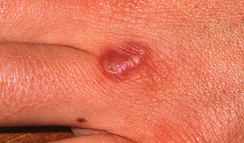
The most common type, localised granuloma annulare, appears in just one or two areas, tends to affect children and young adults, and usually gets better on its own after a few months.

Pink, purple or skin-coloured patches typically appear on the fingers, backs of the hands, feet, ankles or elbows.

They form rings that grow slowly until they’re about 2.5-5cm (1-2 inches) across. As the rings get bigger, they become flatter and more purple in colour before eventually fading.

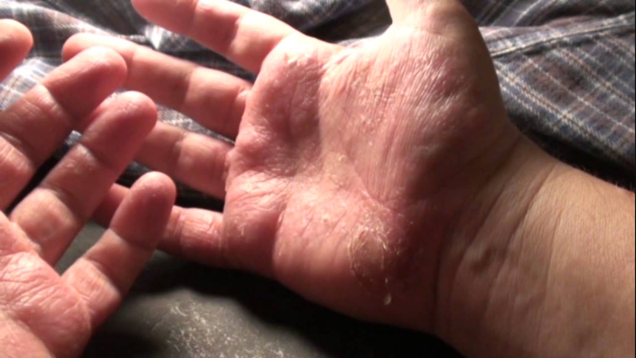
In localised granuloma annulare, the top of the skin feels smooth and, unlike in other skin conditions, such as pityriasis versicolor, ringworm or eczema, it isn’t rough, dry or scaly.

The affected skin also feels firm as a result of inflammation in the middle layer of skin (dermis). There’s no change in the outermost layer of skin (epidermis).

Widespread granuloma annulare

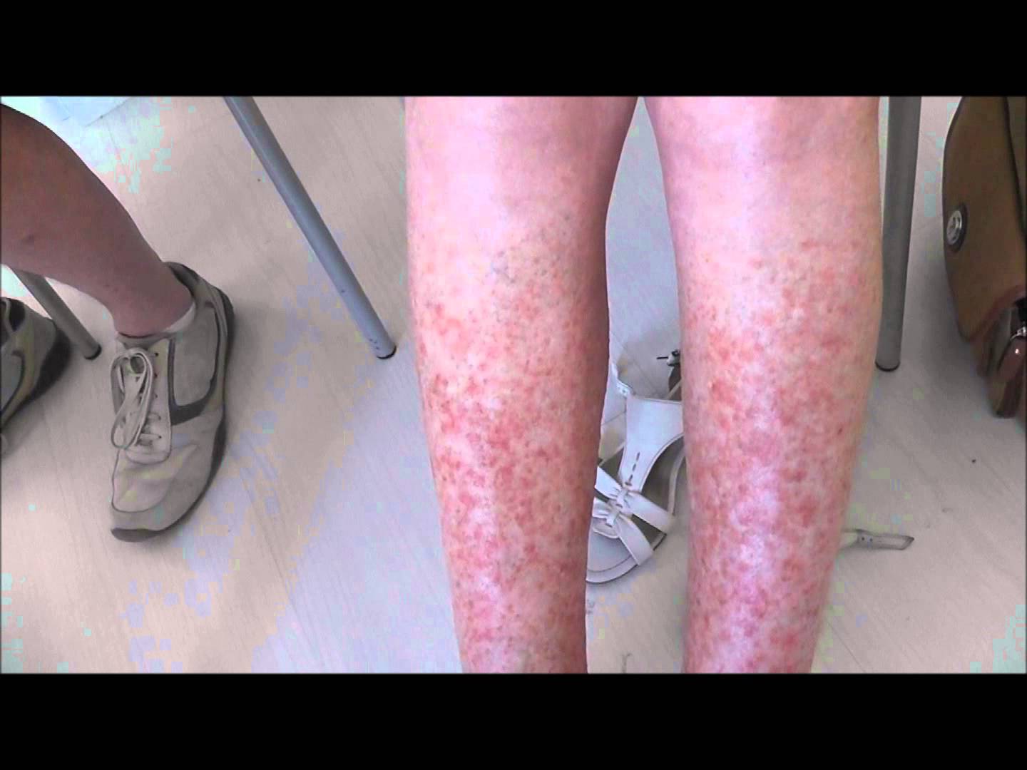
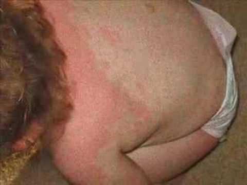
More rarely, you can develop a widespread rash, known as generalised or disseminated granuloma annulare. It usually affects adults.

Large pink, purple or skin-coloured patches appear on a larger portion of the body, including the trunk, arms and legs.

The rash is sometimes made up of small raised spots that form symmetrical rings 10cm (4 inches) or more across. They’re often found in skin folds in the armpits and groin.

Under the skin

Granuloma annulare under the skin usually affects children. One or more firm, rubbery lumps develop under the skin.

They can range from 5mm-4cm (0.2-1.5 inches) in size.

They can appear on the shins, ankles, feet, buttocks, hands, scalp and eyelids.

When to see your GP

See your GP if you have an unexplained rash that doesn’t disappear within a few weeks.

They’ll usually be able to diagnose granuloma annulare from the appearance of your rash.

A skin biopsy may be needed to diagnose the rarer types of granuloma annulare. A small sample of affected skin is taken so it can be studied under a microscope in a laboratory.

A blood glucose test may also be recommended. This is because in rare cases granuloma annulare can be linked to diabetes.

Treating granuloma annulare

Unfortunately, there’s no really effective treatment for granuloma annulare. But in most cases, treatment isn’t necessary because the rash will disappear on its own within a few months to two years.

For more severe cases, you could ask your doctor about the following treatments:

steroid creams or ointments may help, but they must be used carefully because long-term use can thin the skin

cryotherapy (freezing the bumps) may be an option for treating very small patches, but it can be painful and cause permanent scarring

ultraviolet light treatment and powerful medicines (such as steroid tablets, antibiotics, antimalarial medicines, isotretinoin, ciclosporin and dapsone) have been reported to help in individual cases of widespread granuloma annulare, but for most people the symptoms of granuloma annulare don’t justify using these treatments as they all have significant side effects

If you find the appearance of the rash upsetting, you could try using skin camouflage products. Watch this video about skin camouflage and how it can be used to cover marks and scars.

What causes granuloma annulare?

The tissue underneath the top layer of skin (dermis) becomes hypersensitive and inflamed. It’s not clear what causes the skin to react in this way.

Granuloma annulare is harmless and doesn’t affect your overall health. It isn’t caused by allergies and doesn’t run in families. It’s sometimes associated with diabetes, although this is rare.

What’s the best treatment for hand eczema?

Hand eczema

Hand eczema

Hand eczema is a skin condition which affects up to 10% of the UK population. Symptoms include redness, itching, pain, dryness and blisters.

The trial aims to work out which is the most effective hand eczema treatment in people over 18 who have failed to respond to steroid creams. The treatments to be compared in the trial are the tablet treatment Alitretinoin and an ultraviolet light therapy called PUVA, where the hands are exposed to ultra violet (UV) light after they have been soaked in a special solution.

Both of these treatments are used by doctors in standard clinical practice. Some experts also recommend the application of quality eczema creams to prevent it from coming back.  However, there is no clear evidence indicating which is most effective in treating different types of hand eczema. This study will directly compare these two treatments to examine:

– how well the hands heal with the treatment in the short term

– how well the hands heal with the treatment in the longer term

– how long the skin can remain clear once healed

– which of these two treatments is most effective in treating which types of hand eczema.

Kelsey Greet, of Braunstone, Leicester, is a patient participating in the research trial. She said: “I’m a chef, so having hand eczema affects me massively. I struggle to do my job with the pain. I agreed to take part in the study as I hope to regain a bit of normality.”

Dr Graham Johnston, Consultant Dermatologist at Leicester’s Hospitals, said: “We are very pleased to be part of the ALPHA trial team. This is an important NIHR-funded study because it will help identify the most effective treatment for severe hand eczema.”

Dermatology Research Nurse, Sophie Devine, added: “The team is really excited to be part of a clinical research project that can offer our patients further chances to find solutions to what can be a very long-standing and debilitating condition.”

The trial, funded by the National Institute for Health Research, is being led by Leeds’ Clinical Trials Research Unit with colleagues from Bradford Teaching Hospitals NHS Trust.

Dr Miriam Wittmann, Associate Professor in Inflammatory Skin Diseases in the School of Medicine at the University of Leeds, said: “We know how many people really struggle in their everyday lives due to hand eczema, but we do not know definitively which the best treatment to use is.

“This research aims to give patients and clinicians a clear message about which treatment works for them.”

Anyone interested in finding out more about the trial can visit the ALPHA website at http://ow.ly/TJiJN

Polymorphic light eruption – what is it?

Polymorphic light eruption

Polymorphic light eruption

Introduction

Polymorphic light eruption is a fairly common skin rash triggered by exposure to sunlight or artificial ultraviolet (UV) light.

An itchy or burning rash appears within hours, or up to two to three days after exposure to sunlight. It lasts for up to two weeks, healing without scarring.

The rash usually appears on the parts of the skin exposed to sunlight – typically the head and neck, chest and arms (the face isn’t always affected).

Polymorphic light eruption is thought to affect about 10-15% of the UK population.

What does the rash look like?

The rash can take many different forms (known as being “polymorphic”):

most people get crops of 2-5mm pink or red raised spots

some people get blisters that turn into larger, dry red patches (looks a bit like eczema)

less commonly, the skin patches look like targets or bulls-eyes, a bit like the skin rash erythema multiforme

It can be easy to mistake polymorphic light eruption for prickly heat. Prickly heat is caused by warm weather or overheating, rather than sunlight or UV light. The skin in prickly heat doesn’t “harden” or desensitise, as it can do in polymorphic light eruption (see information on desensitisation below).

How big a problem is it?

The skin rash may be a rare occurrence, or may happen every time the skin is exposed to sunlight. It ranges from mild to severe.

Sometimes, as little as 30 minutes of sun exposure is enough to cause the problem, and it can develop even through window glass or thin clothing.

However, for most people with polymorphic light eruption, the rash develops after several hours outside on a sunny day. If further sun is avoided, the rash may settle and go without a trace within a couple of weeks. It may or may not return when skin is re-exposed to sunlight.

If the skin is exposed to more sunlight before the rash has cleared up, it will probably get much worse and spread.

For many affected people, the skin rash appears every spring and remains a problem throughout summer, settling by autumn.

Who’s affected?

Polymorphic light eruption is more common in women than men. It particularly affects those who are fair, although it can affect those with dark skin.

It usually starts from the ages of 20 to 40, although it sometimes affects children.

What is the cause?

Polymorphic light eruption is thought to be caused by UV light altering a substance in the skin, which the immune system then reacts to, resulting in the skin becoming inflamed.

Polymorphic light eruption can run in families and about a fifth of affected people will have an affected relative.

It’s not infectious – there’s no risk of catching polymorphic light eruption from another person.

How is it treated?

There’s no cure for polymorphic light eruption, but careful avoidance of the sun and using sunscreens will help you manage the rash.

Generally, avoid the sun between 11am and 3pm, and wear protective clothing when outdoors (unless you’re hardening your skin; see below). Introduce your skin to sunlight gradually in the spring.

Sunscreen

You may be prescribed sunscreens, which may prevent the rash developing.

Use a sunscreen that is SPF 30 or above with a good UVA rating. Apply it thickly and evenly, reapplying often. Watch a video on how to apply sunscreen.

Steroid creams and ointments

Your GP can prescribe corticosteroid (steroid) cream or ointment, to be applied only when the rash appears. You should apply this sparingly, as often as your GP advises, and never when there’s no rash.

Desensitisation or UV treatment

Sometimes you can increase the resistance of your skin to the sun. This involves visiting a hospital department three times a week for four to six weeks in the spring.

Your skin is gradually exposed to a little more UV light every visit, to try to build up your skin’s resistance. The effects of desensitisation are lost in the winter, so you’ll have to build up your resistance again in the spring.

Hardening or toughening

You may be able to increase the resistance of your skin at home, which is known as “hardening”. This involves going outside for short periods in the spring to build up your resistance.

You might find the time is as short as a few minutes at first, but you may be able to gradually build up to longer times. You’ll have to be careful not to overdo it, but as you understand more about how much light triggers your rash, you’ll be able to judge how long to stay out.

The effects of hardening are lost in the winter, so you’ll have to build up your resistance again in the spring.

Vitamin D

People with polymorphic light eruption are at greater risk of vitamin D deficiency, as a certain amount of sun exposure is needed to make your own vitamin D. Your GP will advise whether you need treatment with vitamin D supplements.

What’s the outlook?

Many people with polymorphic light eruption find the skin complaint improves over years – the skin may harden (become more resistant to sunlight) during the summer, which means more and more sun can be tolerated without the skin reacting. It may even eventually clear up on its own, although this is unusual.

However, hardening of the skin doesn’t always happen, and some people with very sensitive skin may even get the rash in the winter. For these people, it may be a long-term condition to manage with lifestyle changes and creams.

Mastocytosis – what are the signs and symptoms of Mastocytosis?

Mastocytosis causes a wide range of symptoms, which can vary depending on the type of mastocytosis you have.

Cutaneous mastocytosis

Skin lesions are a characteristic of cutaneous mastocytosis. Types of lesions known to occur in cutaneous mastocytosis include:

small areas of skin that change colour (macules)

small firm, raised bumps (papules)

larger raised, red bumps (nodules)

large raised areas of skin noticeable to the touch (plaques)

blisters – which mainly affect young children with mastocytomas (tumours consisting of mast cells) or diffuse cutaneous mastocytosis (a rare form of cutaneous mastocytosis)

Lesions usually develop on the trunk rather than the head, neck and limbs.

The lesions, known as urticaria pigmentosa, are usually yellow-tan to reddish-brown in colour, and can range from 1mm to several centimetres in size.

The number of lesions that develop on the skin can vary widely. For example, it’s possible for only one lesion to develop, or more than 1,000.

Stroking the affected areas of skin can make it swollen, itchy and red over the lesion.

Systemic mastocytosis

If you have systemic mastocytosis, you may develop sudden episodes of symptoms that last for around 15-30 minutes. However, many people don’t have any problems.

The most common symptoms experienced during an episode are:

hot flushing – described as a dry feeling of heat, rather than the sort of wet heat you experience when sweating

a forceful or rapid heartbeat (heart palpitations)

dizziness (lightheadedness)

Less common symptoms during an episode include:

headache

shortness of breath

chest pain

nausea

diarrhoea

Once the episode has passed you’ll probably feel sluggish (lethargic) for several hours.

The episodes are caused by the mast cells suddenly releasing excessive amounts of histamine, usually after you’re exposed to certain triggers.

Triggers known to cause episodes include:

physical factors – such as heat, overheating, cold, fatigue and physical exertion

emotional factors – such as stress and excitement

insect bites or stings – such as flea bites or a wasp sting

infection – such as the cold or flu

alcohol

certain medications – such as ibuprofenaspirin and antibiotics

certain foods – such as cheese, shellfish and spices

Abnormal mast cells in your bone marrow and organs can also cause related symptoms, including:

stomach pain caused by peptic ulcers

loss of appetite

joint pain

weakness

fatigue

changes in mental state – such as confusion, irritability, poor attention span and impaired memory

urinary symptoms – such as needing to pass urine frequently or pain when urinating

In more severe cases of mastocytosis, the following symptoms may occur:

weight loss

swelling of the lymph nodes

swelling of the liver – which can cause jaundice and make you feel lethargic

swelling of the spleen – which can cause tummy (abdominal) and shoulder pain

Low blood pressure (hypotension)

Some people with severe symptoms experience a sudden fall in blood pressure during an attack.

Low blood pressure (hypotension) can trigger a number of associated symptoms, such as:

dizziness

fainting

blurred vision

confusion

general weakness

Severe allergic reaction

If you have systemic mastocytosis or extensive cutaneous mastocytosis, your risk of a severe allergic reaction (anaphylaxis) is increased.

It’s important to look out for the initial symptoms of anaphylaxis, which include:

itchy skin or a raised, red skin rash

swollen eyes, lips, hands and feet

feeling lightheaded or faint

narrowing of the airways, which can cause wheezing and breathing difficulties

abdominal pain, nausea and vomiting

Dial 999 immediately and ask for an ambulance if you think you or someone else is experiencing symptoms of anaphylaxis.

Vitiligo – what are the signs, symptoms, triggers and treatments of vitiligo?

Vitiligo - Michael Jackson had the condition

Vitiligo – Michael Jackson had the condition

Vitiligo is a long-term condition where pale white patches develop on the skin. It’s caused by the lack of melanin, a pigment in the skin.

Vitiligo can affect any area of skin, but most commonly occurs on the face, neck and hands, and in skin creases.

The pale areas of skin are more vulnerable to sunburn, so it’s important to take extra care when in the sun and use a sunscreen with a high sun protection factor (SPF).


Symptoms of vitiligo

The areas most commonly affected by vitiligo include:

the skin around your mouth and eyes

fingers and wrists

armpits

groin

genitals

inside your mouth

It can also sometimes develop where there are hair roots, such as on your scalp. The lack of melanin in your skin can turn the hair in the affected area white or grey.

Vitiligo often starts as a pale patch of skin that gradually turns completely white. The centre of a patch may be white, with pale skin around it. If there are blood vessels under the skin, the patch may be slightly pink, rather than white.

The edges of the patch may be smooth or irregular. They’re sometimes red and inflamed, or there’s brownish discolouration (hyperpigmentation).

Vitiligo doesn’t cause discomfort to your skin, such as dryness, but the patches may occasionally be itchy.

The condition varies from person to person. Some people only get a few small, white patches, but others get bigger white patches that join up across large areas of their skin.

There’s no way of predicting how much skin will be affected. The white patches are usually permanent.

Types of vitiligo

There are two main types of vitiligo:

non-segmental vitiligo

segmental vitiligo

In rare cases, it’s possible for vitiligo to affect your whole body. This is known as universal or complete vitiligo.

Non-segmental vitiligo

In non-segmental vitiligo (also called bilateral or generalised vitiligo), the symptoms often appear on both sides of your body as symmetrical white patches. Symmetrical patches can appear on the:

backs of your hands

arms

skin around body openings, such as the eyes

knees

elbows

feet

Non-segmental vitiligo is the most common type of vitiligo, affecting around nine out of 10 people with the condition.

Segmental vitiligo

In segmental vitiligo (also known as unilateral or localised vitiligo), the white patches only affect one area of your body.

Segmental vitiligo is less common than non-segmental vitiligo, although it’s more common in children. It usually starts earlier and affects three in 10 children with vitiligo.

What causes vitiligo?

Vitiligo is caused by the lack of a pigment called melanin in the skin. Melanin is produced by skin cells called melanocytes, and it gives your skin its colour.

In vitiligo, there aren’t enough working melanocytes to produce enough melanin in your skin. This causes white patches to develop on your skin or hair. It’s not clear exactly why the melanocytes disappear from the affected areas of skin.

Autoimmune conditions

Non-segmental vitiligo (the most common type) is thought to be an autoimmune condition.

In autoimmune conditions, the immune system doesn’t work properly. Instead of attacking foreign cells, such as viruses, your immune system attacks your body’s healthy cells and tissue.

If you have non-segmental vitiligo, your immune system destroys the melanocyte skin cells that make melanin.

Vitiligo is also associated with other autoimmune conditions, such as hyperthyroidism (an overactive thyroid gland), but not everyone with vitiligo will develop these conditions.

Risk factors

You may be at increased risk of developing non-segmental vitiligo if:

other members of your family have it

there’s a family history of other autoimmune conditions – for example, if one of your parents has pernicious anaemia (an autoimmune condition that affects the stomach)

you have another autoimmune condition

you have melanoma (a type of skin cancer) or cutaneous T-cell lymphoma (cancer of the lymphatic system)

you have particular changes in your genes that are known to be linked to non-segmental vitiligo

Neurochemicals

Segmental vitiligo (the less common type) is thought to be caused by chemicals released from the nerve endings in your skin. These chemicals are poisonous to the melanocyte skin cells.

Triggers

It’s possible that vitiligo may be triggered by particular events, such as:

stressful events, like childbirth

skin damage, such as severe sunburn or cuts (this is known as the Koebner response)

exposure to certain chemicals – for example, at work

Vitiligo isn’t caused by an infection and you can’t catch it from someone else who has it.

Diagnosing vitiligo

Your GP will be able to diagnose vitiligo after examining the affected areas of skin.

They may ask whether:

there’s a history of vitiligo in your family

there’s a history of other autoimmune conditions in your family

you’ve injured the affected areas of skin – for example, whether you’ve had sunburn or a severe rash there

you tan easily in the sun, or whether you burn

any areas of skin have got better without treatment, or whether they’re getting worse

you’ve tried any treatments already

Your GP may also ask you about the impact vitiligo has on your life. For example, how much it affects your confidence and self-esteem, and whether it affects your job.

Wood’s lamp

If available, your GP may use an ultraviolet (UV) lamp called a Wood’s lamp to look at your skin in more detail. You’ll need to be in a dark room and the lamp will be held 10-13cm (4-5in) away from your skin.

The patches of vitiligo will be easier to see under the UV light, which will help your GP distinguish vitiligo from other skin conditions, such as pityriasis versicolor (where there’s a loss of pigment due to a fungal infection).

Other autoimmune conditions

As non-segmental vitiligo is closely associated with other autoimmune conditions, you may be assessed to see whether you have any symptoms that could suggest an autoimmune condition, such as:

being tired and lacking energy (signs of Addison’s disease)

being thirsty and needing to urinate frequently (signs of diabetes)

blood test may also be needed to check how well your thyroid gland is functioning.

Treating vitiligo

The white patches caused by vitiligo are usually permanent, although treatment options are available to improve the appearance of your skin.

If the patches are relatively small, skin camouflage cream can be used to cover them up.

In general, combination treatments, such as phototherapy (treatment with light) and medication, give the best results.

Although treatment may help restore colour to your skin, the effect doesn’t usually last. Treatment can’t stop the condition spreading.

Read more about treating vitiligo.

Complications of vitiligo

Vitiligo can sometimes cause other problems.

Because of a lack of melanin, your skin will be more vulnerable to the effects of the sun. Make sure you use a strong sunscreen to avoid sunburn.

Vitiligo may also be associated with problems with your eyes, such as inflammation of the iris (iritis), and a partial loss of hearing (hypoacusis).

Problems with confidence and self-esteem are common in people with vitiligo, particularly if it affects areas of skin that are frequently exposed.