Ichthyosis – what is Ichthyosis?

Ichthyosis is a condition that causes widespread and persistent thick, dry, “fish-scale” skin.

Picture of ichthyosis

There are at least 20 different types of ichthyosis. Some types are inherited at birth and other types are acquired during adulthood.

There’s no cure for ichthyosis, but a daily skincare routine usually keeps the symptoms mild and manageable.

What causes ichthyosis

Most people with ichthyosis have inherited a particular faulty gene from their parent. The signs and symptoms of inherited ichthyosis appear at birth or within the first year of life.

The faulty gene affects the rate at which the skin regenerates – either the shedding of old skin cells is too slow, or the skin cells reproduce at a much faster rate than they can shed old skin. Either way, this causes a build-up of rough, scaly skin.

Ichthyosis vulgaris

Ichthyosis vulgaris is the most common type of inherited ichthyosis, affecting 1 in 250-1,000 people. Signs and symptoms include:

skin may appear normal at birth

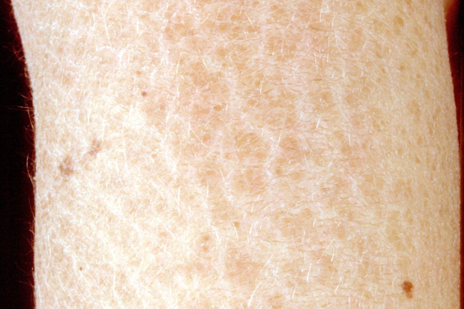
skin gradually becomes dry, rough and scaly, usually before the age of one

the face and the bends of the elbows and knees aren’t usually affected

limbs may develop fine light-grey scales

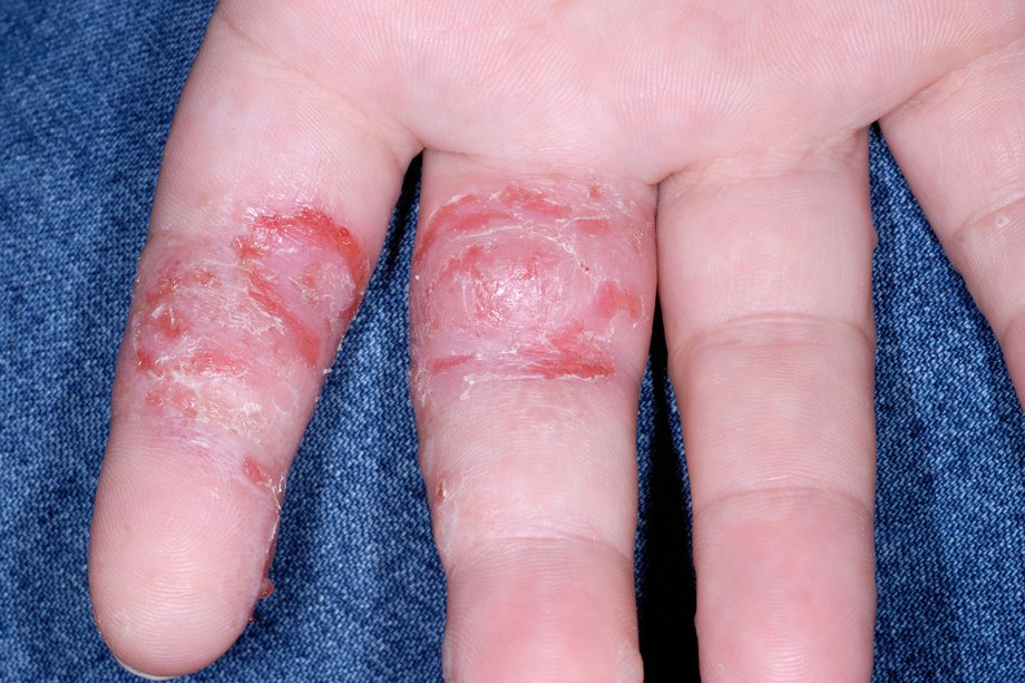
the skin on the palms of the hands and soles of the feet may have more lines than normal and be thickened

the child often also has eczema

symptoms are often worse when it’s cold and dry and improve in warm, humid conditions – this means they may be more noticeable in the winter than the summer


Other types of inherited ichthyosis

Other inherited forms of ichthyosis are very rare and include:

X-linked ichthyosis – only affects males and includes general scaling, particularly over the limbs and trunk (torso)

congenital ichthyosiform erythroderma

harlequin ichthyosis – this is extremely rare, but the scaling is severe and requires intensive care at birth

syndromes that include ichthyosis – such as Netherton’s syndrome or Sjögren-Larsson syndrome

Congenital ichthyosiform erythroderma

Ichthyosis may develop if a baby is born with a shiny yellow membrane (collodion membrane) that sheds within the first week of life.

Once the membrane has shed, one of the following types of ichthyosis can develop:

non-bullous ichthyosiform erythroderma – inflamed scaly skin that affects the entire skin surface

bullous ichthyosiform erythroderma – inflamed scaly skin with fluid-filled blisters that may become infected and produce a foul-smelling skin odour

lamellar ichthyosis – where the skin isn’t as red, but the scales are larger and tighter to the skin

In severe cases of congenital ichthyosiform erythroderma a child may also have drooping lower eyelids (ectropion), mild hair loss and tight skin on the fingers.

Acquired ichthyosis

Acquired ichthyosis tends to develop in adulthood and isn’t inherited. It’s usually associated with another condition, such as:

an underactive thyroid

kidney disease

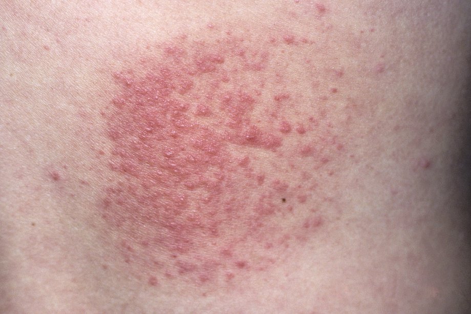
sarcoidosis – a rare condition that causes small patches of red swollen tissue to develop in the body’s organs

Hodgkin lymphoma – a rare type of cancer

HIV infection

Certain medications can also trigger ichthyosis, including some medications used in targeted cancer therapy, such as vemurafenib and protein kinase inhibitors.

Treating ichthyosis

There’s no cure for ichthyosis, but moisturising and exfoliating the skin daily can help prevent dryness, scaling and the build-up of skin cells.

Skincare

Your skin specialist (dermatologist) will be able to prescribe or recommend suitable moisturising treatments (emollients), which may be in the form of a cream, ointment, lotion or bath oil.

You should:

apply emollients to wet skin to trap the moisture – ideally a few minutes after having a bath or shower

gently rub wet skin with a pumice stone to remove some of the thickened skin

brush washed hair to remove scales from your scalp

Other useful exfoliating or moisturising products include lanolin creams, products containing urea, propylene glycol, lactic acid, and other alpha hydroxy acids.

Your dermatologist may also recommend peeling creams, such as salicylic acid, to help exfoliate and moisturise your skin. However, some people may find these products irritate their skin.

Antibiotics or antiseptics may be prescribed to treat skin infections.

Steroid treatments aren’t effective for treating ichthyosis.

Severe ichthyosis

People with severe ichthyosis may need to spend several hours a day caring for their skin.

They may have the following problems:

overheating – as a result of a reduced ability to sweat

limited movement – dry skin can make it too painful to move certain parts of the body

skin infection – after cracking and splitting of the skin

impaired hearing or eyesight – if skin builds up over the ears or eyes

People with severe ichthyosis may be prescribed retinoid tablets (synthetic vitamin A), which reduce the growth of overactive scaly skin. They improve the skin’s appearance, but don’t improve the inflammation or redness.

Vitamin D supplements may also be prescribed.

Outlook

People with mild ichthyosis have a normal lifespan. However, the most severe inherited types can be life threatening.

If you have inherited ichthyosis, you’ll have it for life. Acquired ichthyosis may get better if the underlying cause is identified and treated.

Pompholyx (dyshidrotic eczema)

Pompholyx (dyshidrotic eczema) is a type of eczema that causes tiny blisters to develop across the fingers, palms of the hands and sometimes the soles of the feet.

It can affect people of any age, but it’s most often seen in adults under 40.

Pompholyx can sometimes be confused with similar-looking conditions. See your GP if you have any sort of blistering skin condition.

Signs and symptoms of pompholyx

Pompholyx usually starts as intense itching and burning of the skin on the hands and fingers. The palms and sides of the fingers (and sometimes the soles of the feet) then erupt into tiny itchy blisters that may weep fluid.

Picture of pompholyx

In severe cases, the blisters may be quite large and may spread to the backs of the hands, feet and limbs.

The skin can sometimes become infected. Signs of an infection can include the blisters becoming very painful and oozing pus or becoming covered in a golden crust.

The blisters will usually heal within a few weeks. The skin tends to become dry and crack or peel as it starts to heal.

What causes pompholyx?

It’s not clear exactly what causes pompholyx, but it may be triggered or made worse by:

a fungal skin infection – this may be on the hands or at a distant site from the blisters (such as in between the toes) and will need treating

a reaction to something that has touched your skin – such as certain metals (particularly nickel), detergents, household chemicals, soap, shampoo, cosmetic products or perfume

stress

sweating – pompholyx is more common in spring and summer, in warmer climates, and in people with hyperhidrosis (excessive sweating)

How long does it last?

In many cases, pompholyx will clear up on its own within a few weeks. The treatments below may help relieve your symptoms in the meantime.

Sometimes pompholyx may just occur once and never come back, but it often comes and goes over several months or years. Any of the triggers mentioned above can cause it to flare up again.

Occasionally, pompholyx can be more continuous and difficult to treat.

Treatments for pompholyx

Protecting your skin

You should try to avoid contact with anything that might irritate your skin, including soaps, shampoos and other household chemicals.

Use an emollient as a soap substitute (see below) and wear cotton-lined gloves when you’re at risk of contact with other potentially irritating substances, such as when washing your hair or doing housework.

Don’t burst the blisters – let them heal on their own. If they’re particularly big, your GP may be able to drain them.

Treating the symptoms

The main treatments your GP may recommend to treat the symptoms of pompholyx are similar to those used when treating atopic eczema, including:

emollients (moisturisers) – use these all the time and instead of soap to stop your skin becoming dry

steroid cream – this reduces the inflammation and irritation and helps the skin to heal

Your GP will probably prescribe a strong steroid cream to use for a short period of time, to minimise risk of steroid side effects. You may be advised to wear cotton gloves at night to help the cream sink into the skin.

You can also try:

soaking your hands in a dilute solution of potassium permanganate(1:10,000) for 10 to 15 minutes once or twice a day for up to 5 days

antihistamines to relieve the itching and help you sleep if the itchiness is keeping you awake at night

These treatments are available from pharmacies without a prescription. Your pharmacist can advise whether they’re suitable for you and how you should use them.

Antibiotics may be prescribed if your skin becomes infected.

Specialist treatments

If your pompholyx keeps returning or is severe and doesn’t get better with the above treatments, your GP may refer you to a specialist in treating skin conditions (dermatologist).

A dermatologist may recommend one of the following treatments:

phototherapy – controlled exposure to ultraviolet (UV) light

steroid tablets or very strong steroid cream

immunosuppressant creams or ointments, such as pimecrolimus or tacrolimus

immunosuppressant tablets or capsules, such as ciclosporin or azathioprine

alitretinoin capsules – medication that helps improve severe eczema affecting the hands when other treatments haven’t worked

Similar skin conditions

Conditions that can look similar to pompholyx include:

bullous impetigo – a contagious skin infection that mainly affects children and causes sores and blisters

bullous pemphigoid – a blistering skin condition that tends to affect the elderly

contact dermatitis – a type of eczema caused by skin contact with a substance that causes irritation or an allergic reaction

hand, foot and mouth disease – a viral infection that mainly affects young children, which can cause small blisters to develop on the fingers and palms of the hands

herpetic whitlow (whitlow finger) – an abscess (collection of pus) at the end of the finger that can cause it to become suddenly red, swollen, painful and blistered

pustular psoriasis – an uncommon type of psoriasis that causes pus-filled blisters to appear on your skin

Polymorphic light eruption – common this time of year

Polymorphic light eruption is a fairly common skin rash triggered by exposure to sunlight or artificial ultraviolet (UV) light.

An itchy or burning rash appears within hours, or up to 2 to 3 days after exposure to sunlight. It lasts for up to 2 weeks, healing without scarring.

The rash usually appears on the parts of the skin exposed to sunlight – typically the head, neck, chest and arms. The face is not always affected.

Polymorphic light eruption is thought to affect about 10% to 15% of the UK population.

The rash

A red composed of crops of small red raised spots

The rash can take many different forms (“polymorphic”):

  • most people get crops of 2mm to 5mm pink or red raised spots
  • some people get blisters that turn into larger, dry, red patches – it looks a bit like eczema)
  • less commonly, the skin patches look like targets or bull’s-eyes – it looks a bit like erythema multiforme

Polymorphic light eruption can be easily mistaken for prickly heat. Prickly heat is caused by warm weather or overheating, rather than sunlight or UV light.

The skin in prickly heat does not “harden” or desensitise, as it can do in polymorphic light eruption.

Sunlight exposure

The rash may be a rare occurrence or may happen every time the skin is exposed to sunlight. It ranges from mild to severe.

Sometimes, as little as 30 minutes of sun exposure is enough to cause the problem, and it can even develop through thin clothing or if you’re sitting near a window.

However, for most people with polymorphic light eruption, the rash develops after several hours outside on a sunny day. If further sun is avoided, the rash may settle and disappear without a trace within a couple of weeks. It may or may not return when skin is re-exposed to sunlight.

If the skin is exposed to more sunlight before the rash has cleared up, it will probably get much worse and spread.

For many people with polymorphic skin eruption, the rash appears every spring and remains a problem throughout summer, before settling down by autumn.

Who’s affected?

Polymorphic light eruption is more common in women than men. It particularly affects people who are fair, although it can also affect those with dark skin.

It usually starts between the ages of 20 and 40, although it sometimes affects children.

Causes of polymorphic light eruption

Polymorphic light eruption is thought to be caused by UV light altering a substance in the skin, which the immune system reacts to, resulting in the skin becoming inflamed.

It can run in families – about a fifth of people with the condition have an affected relative.

It’s not infectious, so there’s no risk of catching polymorphic light eruption from another person.

Treatments for polymorphic light eruption

There’s no cure for polymorphic light eruption, but using sunscreens and careful avoidance of the sun will help you manage the rash.

Avoid the sun, particularly between 11am and 3pm when the sun’s rays are at their strongest, and wear protective clothing when outdoors (unless you’re hardening your skin). Introduce your skin to sunlight gradually in the spring.

Sunscreen

You may be prescribed sunscreens to help prevent the rash developing.

Use a sunscreen that is SPF 30 or above with a good UVA rating. Apply it thickly and evenly, reapplying often.

Watch a video on how to apply sunscreen.

Steroid creams and ointments

Your GP can prescribe corticosteroid (steroid) cream or ointment that’s only applied when the rash appears. You should apply it sparingly, as often as your GP advises, and never when there’s no rash.

Desensitisation or UV treatment

It’s sometimes possible to increase the resistance of your skin to the sun. This involves visiting a hospital dermatology department 3 times a week for 4 to 6 weeks in the spring.

Your skin is gradually exposed to a little more UV light every visit to try to build up your skin’s resistance. The effects of desensitisation are lost in the winter, so you’ll have to build up your resistance again in the spring.

Hardening or toughening

You may be able to increase the resistance of your skin at home. This is known as “hardening” and involves going outside for short periods in the spring to build up your resistance.

You might find the time is as short as a few minutes at first, but you may be able to gradually build up to longer times. You’ll have to be careful not to overdo it but, as you begin to understand more about how much light triggers your rash, you’ll be able to judge how long to stay out.

Like desensitisation, the effects of hardening are lost in the winter, so you’ll have to build up your resistance again in the spring.

Vitamin D

People with polymorphic light eruption are at greater risk of vitamin D deficiency, as a certain amount of sun exposure is needed to make your own vitamin D. Your GP will advise whether you need treatment with vitamin D supplements.

Outlook

Many people with polymorphic light eruption find their skin improves over the years. Your skin may harden (become more resistant to sunlight) during the summer, which means more sun can be tolerated without your skin reacting. The rash may even eventually clear up on its own, although this is unusual.

Hardening of the skin does not always happen, and some people with very sensitive skin may even get the rash in the winter. For these people, it may be a long-term condition to manage with lifestyle changes and creams.

Nourish Your Skin With Help From a Dermatologist

We all know how important it is to maintain a healthy diet. But cutting down on the junk food and increasing your intake of vitamins and minerals affects more than just your waistline; it can renew and replenish your skin.

While no food can completely change your complexion by itself, eating right improves your skin just as it improves the rest of your body. So next time you are nourishing your body, do not forget about your largest organ. Your skin spends all day protecting you, isn’t it time you protected it?

The following infographic provides useful tips related to your skin and food for the nourishment of it. Individuals with fair skin are more likely to see problems such as skin cancer and rosacea. So, before starting any skin care regimen, determine your skin tone.

Nourish Your Skin With Help From a Dermatologist

Stevens-Johnson syndrome – What are the signs and symptoms of Stevens-Johnson syndrome?

Stevens-Johnson syndrome is a rare but serious disorder that affects the skin, mucous membrane, genitals and eyes.

The mucous membrane is the soft layer of tissue that lines the digestive system from the mouth to the anus, as well as the genital tract (reproductive organs) and eyeballs.

Stevens-Johnson syndrome is usually caused by an unpredictable adverse reaction to certain medications. It can also sometimes be caused by an infection.

The syndrome often begins with flu-like symptoms, followed by a red or purple rash that spreads and forms blisters. The affected skin eventually dies and peels off.

Stevens-Johnson syndrome is a medical emergency that requires treatment in hospital, often in intensive care or a burns unit.

Treatment aims to identify the underlying cause, control the symptoms and prevent complications.

Erythema multiforme is a similar, but less severe, skin reaction that’s usually caused by infection, particularly herpes viral infections, and a bacteria called mycoplasma that causes chest infections.


Symptoms of Stevens-Johnson syndrome

Skin pain is the most common symptom of Stevens-Johnson syndrome.

Flu-like symptoms are also usually present during the initial stages, and may include:

feeling generally unwell

a high temperature (fever) of 38C (100.4F) or above

headache

joint pain

cough

After a few days a rash appears, which may look like a target – darker in the middle and lighter around the outside (see below). The rash isn’t usually itchy, and spreads over a number of hours or days. Large blisters then develop on the skin, which after bursting leave painful sores.

Facial swelling and swollen lips covered in crusty sores are common features of Stevens-Johnson syndrome. The mucous membranes inside your mouth, throat, eyes and genital tract may also become blistered and ulcerated. This can make swallowing painful and lead to serious problems such as dehydration.

The surface of the eyes can also sometimes be affected, which if not treated quickly can cause corneal ulcers and vision problems

Causes of Stevens-Johnson syndrome

In children, Stevens-Johnson syndrome is usually triggered by a viral infection, such as

mumps

flu

herpes-simplex virus, which causes cold sores

Coxsackie virus, which causes Bornholm disease

Epstein-Barr virus, which causes glandular fever

Less commonly, bacterial infections can also trigger the syndrome.

In adults, Stevens-Johnson syndrome is often caused by an adverse reaction to medication. The medications associated with a high risk of Stevens-Johnson syndrome are:

allopurinol

carbamazepine

cotrimoxazole and other anti-infective sulfonamides – including sulfasalazine, sulfadiazine, sulfadoxine and sulfafurazole

lamotrigine

nevirapine

certain types of non-steroidal anti-inflammatory drugs (NSAIDs) – including meloxicam, piroxicam and tenoxicam

phenobarbital

phenytoin

sertraline

As a result of the associated risk, a thorough evaluation of the expected benefits of treatment is required when prescribing the above medications. Doctors are advised to carefully consider using these medications as first-line treatments, particularly if there are safer alternatives.

It’s important to emphasise that Stevens-Johnson syndrome is rare, and the overall risk of getting the syndrome is low, even for people using “high risk” medications (one in 1,000 to one in 100,000).

Risk factors

Risk factors for Stevens-Johnson syndrome may include:

viral infections – such as herpes, hepatitis, viral pneumonia or HIV

a weakened immune system – as a result of HIV or AIDS, autoimmune conditions, such as lupus, or certain treatments, such as chemotherapy and organ transplants

a previous history of Stevens-Johnson syndrome – if the syndrome was previously caused by medication, you’re at risk of it reoccurring if you take the same medication again, or medications from the same family of medications

a family history of Stevens-Johnson syndrome – if a close family member has had the syndrome, your risk of getting it may be increased

Specific genes have also been identified that increase the risk of Stevens-Johnson syndrome among certain groups of people.

For example, Chinese people with the HLA B1502 gene have experienced Stevens-Johnson syndrome after taking carbamazepine, and allopurinol has also triggered the syndrome in Chinese people with the HLA B1508 gene.

Diagnosing Stevens-Johnson syndrome

Stevens-Johnson syndrome should be diagnosed by a dermatologist (skin specialist). A diagnosis is often based on a combination of your:

symptoms

physical examination

medical history (including any medication you’ve recently taken)

To confirm the diagnosis, a small sample of skin may be removed (biopsy) so that it can be tested in a laboratory.

Treating Stevens-Johnson syndrome

If Stevens-Johnson syndrome is suspected, you or your child will be immediately referred to hospital for treatment. Without treatment, the symptoms can become very severe and be life-threatening.

Severe cases of Stevens-Johnson syndrome may need to be treated in an intensive care unit (ICU) or burns unit.

The first step is to stop taking any medications that may be causing Stevens-Johnson syndrome. However, it can sometimes be difficult to determine which medication is causing it, so stopping all non-essential medications may be recommended.

Treatment for relief of symptoms while in hospital may include:

cool, moist compresses held against the skin – dead skin may be gently removed and a sterile dressing placed over the affected area

regularly applying a plain (unscented) moisturiser to the skin

replacement fluids – you may receive fluids and nutrition through a tube that’s passed through your nose and into your stomach (a nasogastric tube)

mouthwashes containing anaesthetic and/or antiseptic – to temporarily numb your mouth and make swallowing easier

a short course of corticosteroid tablets (topical corticosteroids) to control skin inflammation (only on specialist advice)

antibiotics – if blood poisoning (sepsis) is suspected

eyedrops or eye ointment – for eye-related symptoms

Once the cause of Stevens-Johnson syndrome has been identified and successfully treated (in the case of an infection), or stopped (in the case of medication), the skin reaction will stop. New skin may start to grow after a few days.

However, the length of time it takes to recover from Stevens-Johnson syndrome will depend on how severe it is, and it can sometimes take many weeks or months to fully recover.

If the cause was an adverse reaction to medication, you’ll need to avoid that medication and possibly other similar medications for the rest of your life. The doctor treating you will be able to advise you further about this.

Complications of Stevens-Johnson syndrome

As Stevens-Johnson syndrome severely affects the skin and mucous membranes, it can cause a number of complications. These include:

skin changes – when your skin grows back it may be uneven in colour; less commonly, scarring may occur

secondary skin infection (cellulitis) – which can lead to further, serious problems such as blood poisoning (sepsis)

problems with internal organs – organs can become inflamed – for example, the lungs (pneumonia), heart (myocarditis), kidneys (nephritis) or liver (hepatitis); the oesophagus may also become narrowed and scarred (oesophageal stricture)

eye problems – the rash can cause problems with your eyes which, in mild cases, may be irritation and dry eyes, or in severe cases may result in corneal ulceration, uveitis (inflammation of the uvea, which is the middle layer of the eye) and, possibly, blindness

Problems with the sexual organs, such as vaginal stenosis (narrowing of the vagina caused by a build-up of scar tissue), and scarring of the penis, is also a possible complication of Stevens-Johnson syndrome.

Preventing Stevens-Johnson syndrome

If Stevens-Johnson syndrome has been caused by an adverse reaction to a medication, you’ll need to avoid taking this medication and other similar medications. Other family members may also want to avoid using the medication in case there’s a genetic susceptibility within your family.

If you’ve had Stevens-Johnson syndrome in the past, and your doctor thinks you’re at risk of getting it again in the future, you’ll be warned to look out for the symptoms.

If you’re of Chinese, southeast Asian or Indian descent, genetic testing may be recommended before taking medications, such as carbamazepine and allopurinol, that are known to have an associated risk of causing Stevens-Johnson syndrome. Testing will help determine whether you carry the genes (HLA B1502 and HLA B1508) that have been associated with the syndrome when taking these medications.