How a common food ingredient can take a wrong turn, leading to rheumatoid arthritis

# #

We need tryptophan to survive, but bacteria can break it down into an inflammatory chemical; a CU Department of Medicine faculty member explores how that process works in new research.

#

University of Colorado Department of Medicine faculty member says she and her colleagues have identified how bacteria in the digestive system can break down tryptophan in the diet into an inflammatory chemical that primes the immune system towards arthritis.

The research was co-authored by Kristine Kuhn, MD, PhD, Scoville Endowed Chair and head of the CU Division of Rheumatology. Several of her division colleagues collaborated on the paper, which was published in February in the Journal of Clinical Investigation.

#

Tryptophan is an essential amino acid found in many protein-rich foods, including meats, fish, dairy products, and certain seeds and nuts. It is also used in the body to help produce proteins, muscles, enzymes, and neurotransmitters—the nervous system’s chemical messengers. The body doesn’t make tryptophan; we get it from our diet.

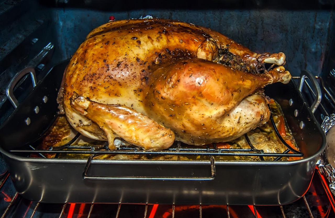
Many people think of tryptophan as the ingredient in turkey that supposedly makes us sleepy after a Thanksgiving feast. In fact, researchers say that although tryptophan plays a role in helping to regulate the sleep cycle, the amount that’s in turkey probably isn’t a significant cause of post-dinner drowsiness.

Cause and effect

Kuhn and her associates set out to learn how a substance that often is a force for good in the body is converted into a pathway to inflammatory diseases such as rheumatoid arthritis, which affects about 1% of the population. It can cause painful swelling of the hands and feet, and joint deformities if left untreated.

“It’s been known that the microbiome – the bacteria in our gut – can break down tryptophan into byproducts. Some of those byproducts are anti-inflammatory, but we’ve also associated some inflammatory causes of those products,” Kuhn says. “We’re the first to highlight which products are contributing to inflammation, and how they are doing that.”

She says the new research “builds upon some observations we had in patients with spondyloarthritis – not quite rheumatoid arthritis, but a closely related condition – where we found that changes in the microbiome were associated with increased production of these products called indoles, which are what bacteria make from tryptophan.” Similar changes were observed in arthritis studies involving mice, she says.

“We put mice on antibiotics to wipe out their microbiome, and they didn’t get arthritis, and they didn’t have indole,” she says. “So we said, OK, what if they do have a microbiome and we put them on a diet with little tryptophan? The microbiome can’t break down tryptophan into indole, and the mice didn’t get arthritis. So two different ways, we showed that it’s tryptophan that’s broken down by the microbiome into indole.”

Inflammatory flags

So how does that work? “We found that when indole is present, the mice start to develop autoreactive T-cells that are more inflammatory. They have less of those regulatory T-cells that help maintain balance in the immune system, and they start to develop antibodies that are more pathogenic. We found that the antibodies had flags for being more inflammatory when indole was present.”

The paper concludes that “blockade of indole generation may present a unique therapeutic pathway” for rheumatoid arthritis and spondyloarthritis. That’s all about finding the right path for the body’s tryptophan, Kuhn says.

“If tryptophan hits our body’s cells, it tends to go get broken down into anti-inflammatory products versus when it hits the bacterial cells and goes more inflammatory. The ways we think about how this could lead to therapies are: How do you keep that balance tipped so that tryptophan goes towards that anti-inflammatory pathway? How can you manipulate intestinal bacteria to tip that balance? That’s where we’re interested in going in the future.”

Does Kuhn’s research suggest we should be eating differently? “I get asked that a lot,” she says. “A diet that’s rich in plant-based fibers and lean meats – this whole Mediterranean diet – seems to push the microbiome into a healthier state, so that you are getting the anti-inflammatory properties of tryptophan, whereas the typical western diet seems to go more toward the inflammatory pathway.”

As for other ways to protect against arthritis, Kuhn says that through research by her Division of Rheumatology colleagues, “we have started to understand the at-risk stage, where we can actually identify people who are likely to progress to rheumatoid arthritis within the next few years based on blood markers. There’s some data that suggests we could intervene during that period and prevent disease, but we’re not quite sure yet what are the right ways to intervene.”

RA and Lupus – Scientists reveal how our cells’ leaky batteries make us sick.

#

Scientists reveal how our cells’ leaky batteries are making us sick

#

“Now that we are beginning to understand how this inflammation starts, we might be able to prevent this process, with the ultimate goal of limiting inflammation and treating disease,” said researcher Laura E. Newman, PhD. CREDIT Courtesy Newman lab

Researchers have discovered how “leaky” mitochondria – the powerhouses of our cells – can drive harmful inflammation responsible for diseases such as lupus and rheumatoid arthritis. Scientists may be able to leverage the findings to develop better treatments for those diseases, improve our ability to fight off viruses and even slow ageing.

#

The new discovery reveals how genetic material (mitochondria) can escape from our cellular batteries and prompt the body to launch a damaging immune response. By developing therapies to target this process, doctors may one day be able to stop the harmful inflammation and prevent its toll on our bodies.

“When mitochondria don’t correctly replicate their genetic material, they try to eliminate it. However, if this is happening too often and the cell can’t dispose of all of it, it can cause inflammation, and too much inflammation can lead to disease, including autoimmune and chronic diseases,” said researcher Laura E. Newman, PhD, of the University of Virginia School of Medicine. “Now that we are beginning to understand how this inflammation starts, we might be able to prevent this process, with the ultimate goal of limiting inflammation and treating disease.”

#

Powering Inflammation

Mitochondria have their own set of genetic material, separate from the DNA that serves as the operating instructions for our cells. Scientists have known that this mitochondrial DNA, mtDNA, can escape into our cells and cause inflammation. But exactly what caused this has been a mystery until now.

“We knew that mtDNA was escaping mitochondria, but how was still unclear,” said Gerald Shadel, PhD, director of the San Diego-Nathan Shock Center of Excellence in the Basic Biology of Aging at the Salk Institute. “Using imaging and cell biology approaches, we’re able to trace the steps of the pathway for moving mtDNA out of the mitochondria, which we can now try to target with therapeutic interventions to prevent the resulting inflammation hopefully.”

Shadel and Newman, then a postdoctoral researcher in Shadel’s lab, and their collaborators used sophisticated imaging techniques to determine what was happening inside the leaky mitochondria. They found that a malfunction in mtDNA replication triggered the leak. This caused the accumulation of protein masses caused by nucleoids.

To try to fix this problem, the cell containing the faulty mitochondrion begins to export the excess nucleoids to its cellular trash bins. But the trash bins, called endosomes, can become overwhelmed by the volume of debris, the scientists found. These overburdened endosomes respond by releasing mtDNA into the cell – in short, the trash can overflows.

“We had a huge breakthrough when we saw that mtDNA was inside of a mysterious membrane structure once it left mitochondria. After assembling all of the puzzle pieces, we realized that structure was an endosome,” Newman said. “That discovery eventually led us to the realization that the mtDNA was being disposed of and, in the process, some of it was leaking out.”

The cell responds to this hazardous waste spill by flagging the nucleoids as foreign DNA, like a virus, and launches an immune response that results in harmful inflammation, the scientists determined.

“Using our cutting-edge imaging tools for probing mitochondria dynamics and mtDNA release, we have discovered an entirely novel release mechanism for mtDNA,” said researcher Uri Manor, PhD, former director of the Waitt Advanced Biophotonics Core at Salk and current assistant professor at UC San Diego. “There are so many follow-up questions we cannot wait to ask, like how other interactions between organelles control innate immune pathways, how different cell types release mtDNA, and how we can target this new pathway to reduce inflammation during disease and aging.”

Newman will continue to seek these answers in her new role at the UVA School of Medicine’s Department of Cell Biology. “We want to understand the physiological and disease contexts where this process can become activated,” she said. “For example, many viruses attack mitochondria during infection, so we will be testing whether mitochondria purposely use this pathway to sound the alarm against invading viruses, and whether over-reliance on this pathway to fight off infection can later trigger chronic diseases.”

Findings Published

The Ultimate Diet Guide for Managing Rheumatoid Arthritis: A Comprehensive Approach

Join Dr. Diana Girnita, a double board-certified Rheumatologist and Internal Medicine specialist, for a live YouTube lecture on “The Ultimate Diet Guide for Managing Rheumatoid Arthritis: A Comprehensive Approach.” Let us answer these questions: What are the worst foods for rheumatoid arthritis? The link between your gut and rheumatoid arthritis What are the best foods for rheumatoid arthritis? Throughout this video, we will explore various dietary recommendations scientifically proven to alleviate symptoms of rheumatoid arthritis. From incorporating anti-inflammatory foods to avoiding potential trigger foods, Dr. Girnita will explain how certain dietary choices can help minimize joint pain, swelling, and morning stiffness. You will learn about foods with these invaluable properties and how they can actively support your body’s healing and anti-inflammatory processes.

https://youtube.com/watch?v=HPK4ZfwKz_k%3Fsi%3DCNZiJHxVXkNzEODc

New treatment to reverse inflammation and arterial blockages in rheumatoid arthritis announced

Researchers from Queen Mary University of London have found that the molecule RvT4 enhances the body’s natural defences against atherosclerosis (hardening of the arteries) in patients with rheumatoid arthritis. 

Studies in mice undertaken by researchers from Queen Mary University of London’s William Harvey Research Institute and Centre for Inflammation and Therapeutic Innovation, and funded by the European Research Council (ERC) and Barts Charity, show that increasing levels of the RvT4 molecule in the body improves the ability of the body’s own defence mechanisms [macrophages] to reduce local inflammation and remove blockages in blood vessels. This breakthrough in understanding the processes involved could lead to better treatments for people who have rheumatoid arthritis (RA), and who are at higher risk of developing cardiovascular disease.  

Rheumatoid arthritis (RA) is the most common form of inflammatory arthritis in the UK and affects around 1% of the population. Approximately 10,000 people receive a diagnosis of RA every year. Alongside the more widely-known symptoms of joint inflammation, people with the condition are also twice as likely as others to develop blood vessel disease. This can lead to serious complications and an increased risk of premature death. 

One type of blood vessel disease seen in people with RA is atherosclerosis, which is caused by a build-up of fatty material called ‘plaque’ along the artery walls. This build-up causes the arteries to harden and narrow, making it more difficult to circulate blood around the body. These blockages can also break free, causing heart attacks and strokes. Understanding the reasons why RA patients are at increased risk of these cardiovascular problems is critical in developing better treatments for this group and others. 

To better understand the causes of blood vessel disease in patients with RA, researchers explored the role of a group of molecules called 13-series resolvins (RvTs). In experimental arthritis, the levels of one of these molecules, RvT4, are markedly reduced, a phenomenon that is associated with a higher degree of blood vessel disease. This study was designed to explore why this might be the case. 

The findings 

The study found that treating arthritic mice with RvT4 reduced blood vessel inflammation by re-programming macrophages – a group of white blood cells that accumulate in the diseased vessels – to release stored lipids. Researchers observed that these lipids were preventing the macrophage from carrying out their usual work of clearing dead cells and reducing localised inflammation in blood vessels. Once freed of their lipid burden, the macrophages were able to move and work much more effectively to reduce the causes of atherosclerosis. The observation that RvT4 restores protective macrophage biological activities is an exciting finding.   

RA patients also often present with metabolic dysfunction and this is thought to exacerbate vascular disease. The study found that administration of RvT4 to mice engineered to develop characteristics of metabolic dysfunction, advanced atherosclerosis, and arthritis led to an overall decrease in lipoprotein-associated cholesterol in plasma and an increase in the ratio of HDL-associated cholesterol to total cholesterol. 

Study shows why women are at greater risk of autoimmune disease.

Women and pain

omewhere between 24 and 50 million Americans have an autoimmune disease, a condition in which the immune system attacks our own tissues. As many as 4 out of 5 of those people are women.

Rheumatoid arthritis, multiple sclerosis and scleroderma are examples of autoimmune disorders marked by lopsided female-to-male ratios. The ratio for lupus is 9 to 1; for Sjogren’s syndrome, it’s 19 to 1.

Stanford Medicine scientists and their colleagues have traced this disparity to the most fundamental feature differentiating biological female mammals from males, possibly paving the way for a better way to predict autoimmune disorders before they develop.

“As a practising physician, I see a lot of lupus and scleroderma patients because those autoimmune disorders manifest in the skin,” said Howard Chang, MD, PhD, dermatology professor and genetics professor and a Howard Hughes Medical Institute investigator. “The great majority of these patients are women.”

Chang, the Virginia and D.K. Ludwig Professor in Cancer Research and director of the RNA Medicine Program, is the senior author of the study, to be published Feb. 1 in Cell. Basic life research scientist Diana Dou, PhD, is its lead author.

The silence of the second X

Women have too much of a good thing: It’s called the X chromosome.

Throughout the mammalian kingdom, biological sex is determined by the presence of two X chromosomes in every female cell. Male cells pack just one X chromosome, paired with a much shorter one designated the Y chromosome.

The stubby Y chromosome contains only a handful of active genes. It’s quite possible to live a full life without a Y chromosome. In fact, more than half of the people on Earth — women — lack Y chromosomes and do just fine. But no mammalian male or female cell can survive without at least one copy of the X chromosome, which holds many hundreds of active protein-specifying genes.

Still, having two X chromosomes risks the production, in every female cell, of twice the amount of the myriad proteins specified by the X but not the Y chromosome. Such massive overproduction of so many proteins would be lethal.

Nature has devised a clever, if complicated, workaround called X-chromosome inactivation. Early in embryogenesis, each cell in the nascent female mammal decides to shut down the activity of one or the other of its two X chromosomes. Once that decision is made, it’s handed down to these cells’ progeny in the developing fetus. That way, the same amount of each X-chromosome-specified protein is produced in a female cell as in a male cell.

As the researchers discovered, X-chromosome inactivation can lead to autoimmune disorders, but other factors can also cause these disorders — which is why men sometimes develop them.

The great equalizer

X-chromosome inactivation is achieved courtesy of a molecule called Xist. The gene for Xist is present on all X chromosomes, including the single one male cells have. But Xist itself is produced only when the X chromosome that its gene resides on is one of a matched XX pair — and is produced and deployed on only one pair member.

Xist consists of RNA, a substance best known for being a simple-minded messenger that shuttles genes’ instructions for making proteins to the intracellular machines that make them. Yet RNA can do a whole lot more than schlep genetic information. There are as many different kinds of so-called long noncoding RNA (lncRNA) molecules — lengthy RNA stretches that don’t carry instructions for making proteins — as there are of the protein-encoding RNA variety. These lncRNA molecules can park themselves on stretches of chromosomes and change the likelihood that the cellular machinery charged with reading the genes in those locations will do so.

Xist, a type of lncRNA, is much longer than most. Xist coats long sections of one of a female mammalian cell’s two X chromosomes — but always just one — cutting that chromosome’s output to zero or close to it. The other X chromosome, left undisturbed, pumps out just enough RNA-encoded instructions to keep the cell humming.

But Xist’s nestling into the extra X chromosome generates odd combinations of lncRNA, proteins that bind to it, other proteins that bind to those proteins, and DNA some of those proteins cling to. These complexes can trigger a strong immune response, Chang and his colleagues have learned.

In 2015, Chang’s group identified close to 100 proteins that either bound to Xist or that bound to those proteins, collectively enabling this molecule to lay anchor along gene-specifying regions of the X chromosome.

Inspecting this Xist “parts list,” Chang realized that many of Xist’s collaborator proteins were known to be associated with autoimmune disorders. Might the RNA-protein-DNA complexes generated in the course of X-chromosome inactivation be triggering the notoriously high rate of autoimmunity in women compared with men? That question was the impetus for the new study.

What if males made Xist?

To eliminate possible competing causes such as female hormonal action or aberrant protein production by the supposedly silenced second X chromosome, the researchers tossed the Xist ball into the male court. They sewed the gene for Xist into the genomes of two different strains of male lab mice. One strain is quite susceptible to autoimmune symptoms mimicking lupus, with females more susceptible than males. The other is resistant to it.

The inserted Xist gene had been modified in two ways. It could be turned on or off by chemical means, pumping out Xist only when the scientists wanted it to. The Xist gene was also tweaked slightly so that its RNA product would no longer silence the genes of the male mouse’s chromosome into which it was stitched.

Merely inserting that modified Xist gene had no noticeable effect on the mice. But the Xist produced from the inserted gene, once that gene was activated, still formed characteristic complexes with almost all the proteins found earlier to be collaborating closely with Xist.

Now, the scientists could ask: Is a bioengineered male mouse that’s been coaxed to produce Xist more prone to autoimmunity than a normal male mouse, which never produces it, or than a male in whom the gene for Xist has been inserted but not activated?

By injecting an irritant known to induce a lupus-like autoimmune condition in the susceptible mouse strain, the investigators could compare its effect on males who made Xist with its effect on normal males, who made none.

In these susceptible mice, males in which the Xist gene was activated developed lupus-like autoimmunity at a rate approaching that of females — and considerably more so than non-bioengineered males.

The absence of autoimmunity in some female or Xist-activated male mice in the susceptible strain showed that not just activation of Xist but also some kind of tissue-damaging stress (caused, in this case, by injection of the irritant) is required to get the autoimmunity ball rolling.

In the autoimmune-resistant strain, activating Xist in bioengineered male mice wasn’t enough to induce autoimmunity — as might be predicted by the fact that in this strain even females seldom develop autoimmunity. That suggests that not only Xist activation but also an appropriate genetic background is necessary for autoimmunity to develop.

These constraints on autoimmunity are fortunate, because if there were none all women might be more susceptible to develop immunity, Chang noted.

Toward a better autoimmunity-screening panel

An early step in the development of autoimmunity is the appearance of autoantibodies: antibodies targeting one’s own tissues or cell products. Autoantibodies to the contents of cell nuclei are called anti-nuclear antibodies. Close examination of blood samples from about 100 patients with autoimmunity showed the presence of autoantibodies to many of the complexes associated with Xist. Some of these autoantibodies were specific to one or another autoimmune disorder, indicating their potential utility in identifying particular emergent autoimmune disorders before symptoms develop. Autoantibodies to still other Xist-associated proteins spanned several disorders, designating them as possible common markers of autoimmunity.

“Every cell in a woman’s body produces Xist,” Chang said. “But for several decades, we’ve used a male cell line as the standard of reference. That male cell line produced no Xist and no Xist/protein/DNA complexes, nor have other cells used since for the test. So, all of a female patient’s anti-Xist-complex antibodies — a huge source of women’s autoimmune susceptibility — go unseen.”