Decoding a direct dialogue between the gut microbiota and the brain

Decoding a direct dialog between the gut microbiota and the brain

Decoding a direct dialog between the gut microbiota and the brain CREDIT © Institut Pasteur / Pascal Marseaud

Gut microbiota by-products circulate in the bloodstream, regulating host physiological processes including immunity, metabolism and brain functions. Scientists from the Institut Pasteur (a partner research organization of Université Paris Cité), Inserm and the CNRS have discovered that hypothalamic neurons in an animal model directly detect variations in bacterial activity and adapt appetite and body temperature accordingly. These findings demonstrate that a direct dialog occurs between the gut microbiota and the brain, a discovery that could lead to new therapeutic approaches for tackling metabolic disorders such as diabetes and obesity. The findings are due to be published in Science on April 15, 2022.

The gut is the body’s largest reservoir of bacteria. A growing body of evidence reveals the degree of interdependence between hosts and their gut microbiota, and emphasizes the importance of the gut-brain axis. At the Institut Pasteur, neurobiologists from the Perception and Memory Unit (Institut Pasteur/CNRS)[1], immunobiologists from the Microenvironment and Immunity Unit (Institut Pasteur/Inserm), and microbiologists from the Biology and Genetics of the Bacterial Cell Wall Unit (Institut Pasteur/CNRS/Inserm)[2] have shared their expertise to investigate how bacteria in the gut directly control the activity of particular neurons in the brain.

The scientists focused on the NOD2 (nucleotide oligomerization domain) receptor which is found inside of mostly immune cells. This receptor detects the presence of muropeptides, which are the building blocks of the bacterial cell wall. Moreover, it has previously been established that variants of the gene coding for the NOD2 receptor are associated with digestive disorders, including Crohn’s disease, as well as neurological diseases and mood disorders. However, these data were insufficient to demonstrate a direct relationship between neuronal activity in the brain and bacterial activity in the gut. This was revealed by the consortium of scientists in the new study.

Using brain imaging techniques, the scientists initially observed that the NOD2 receptor in mice is expressed by neurons in different regions of the brain, and in particular, in a region known as the hypothalamus. They subsequently discovered that these neurons’ electrical activity is suppressed when they come into contact with bacterial muropeptides from the gut. “Muropeptides in the gut, blood and brain are considered to be markers of bacterial proliferation,” explains Ivo G. Boneca, Head of the Biology and Genetics of the Bacterial Cell Wall Unit at the Institut Pasteur (CNRS/Inserm). Conversely, if the NOD2 receptor is absent, these neurons are no longer suppressed by muropeptides. Consequently, the brain loses control of food intake and body temperature. The mice gain weight and are more susceptible to developing type 2 diabetes, particularly in older females.

In this study, the scientists have demonstrated the astonishing fact that neurons perceive bacterial muropeptides directly, while this task was thought to be primarily assigned to immune cells. “It is extraordinary to discover that bacterial fragments act directly on a brain center as strategic as the hypothalamus, which is known to manage vital functions such as body temperature, reproduction, hunger and thirst,” comments Pierre-Marie Lledo, CNRS scientist and Head of the Institut Pasteur’s Perception and Memory Unit.

The neurons thus appear to detect bacterial activity (proliferation and death) as a direct gauge of the impact of food intake on the intestinal ecosystem. “Excessive intake of a specific food may stimulate the disproportionate growth of certain bacteria or pathogens, thus jeopardizing intestinal balance,” says Gérard Eberl, Head of the Microenvironment and Immunity Unit at the Institut Pasteur (Inserm).

The impact of muropeptides on hypothalamic neurons and metabolism raises questions on their potential role in other brain functions, and may help us understand the link between certain brain diseases and genetic variants of NOD2. This discovery paves the way for new interdisciplinary projects at the frontier between neurosciences, immunology and microbiology, and ultimately, for new therapeutic approaches to brain diseases and metabolic disorders such as diabetes and obesity.


[1] This research unit is also known as the “Genes, Synapses and Cognition Laboratory” (Institut Pasteur/CNRS).
Paris Brain Institute (CNRS/Inserm/Sorbonne Université/AP-HP) also contributed to these findings.

[2] The CNRS unit’s name is the “Integrative and Molecular Microbiology Unit” and the Inserm unit’s name is the “Host-Microbe Interactions and Pathophysiology Unit” (Institut Pasteur/CNRS/Inserm).
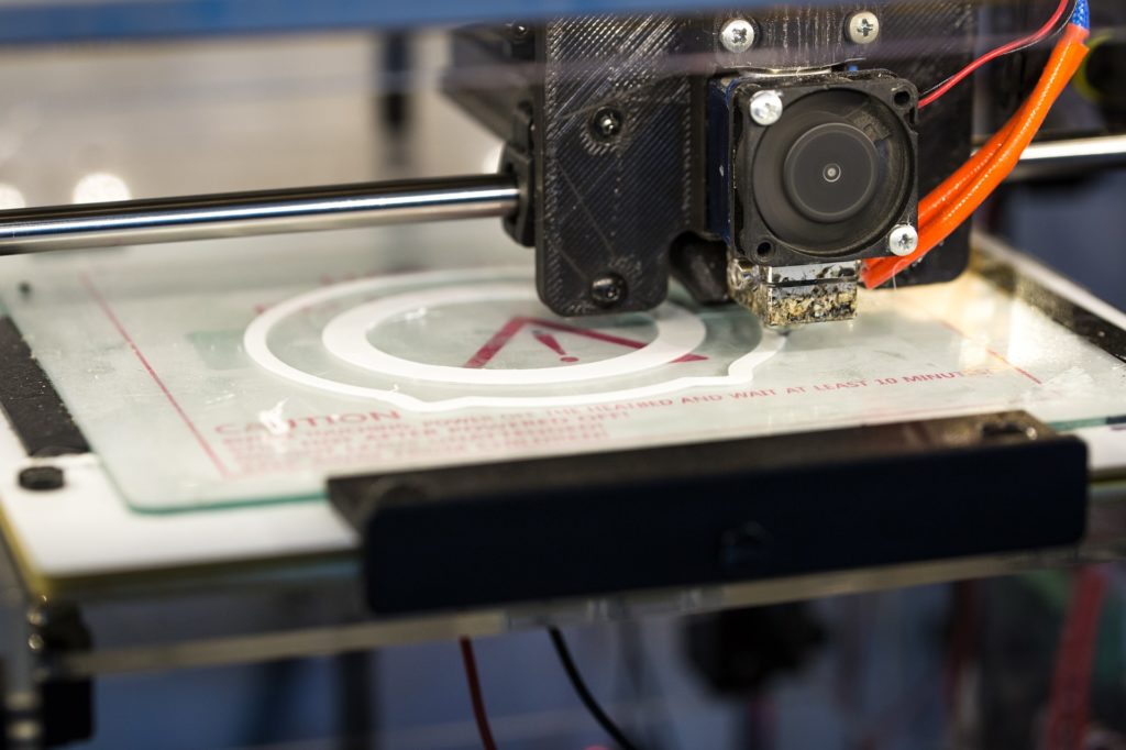
New 3D Printing technique: a game-changer for medical testing devices

New 3D Printing technique: a game changer for medical testing devices


Microfluidic devices are compact testing tools made up of tiny channels carved on a chip, which allow biomedical researchers to test the properties of liquids, particles and cells at a microscale. They are crucial to drug development, diagnostic testing and medical research in areas such as cancer, diabetes and now COVID-19. However, the production of these devices is very labour-intensive, with minute channels and wells that often need to be manually etched or moulded into a transparent resin chip for testing. While 3D printing has offered many advantages for biomedical device manufacturing, its techniques were previously not sensitive enough to build layers with the minute detail required for microfluidic devices. Until now.

Researchers at the USC Viterbi School of Engineering have now developed a highly specialized 3D printing technique that allows microfluidic channels to be fabricated on chips at a precise microscale not previously achieved. The research, led by Daniel J. Epstein Department of Industrial and Systems Engineering Ph.D. graduate Yang Xu and Professor of Aerospace and Mechanical Engineering and Industrial and Systems Engineering Yong Chen, in collaboration with Professor of Chemical Engineering and Materials Science Noah Malmstadt and Professor Huachao Mao at Purdue University, was published in Nature Communications.

The research team used a type of 3D printing technology known as vat photopolymerization, which harnesses light to control the conversion of liquid resin material into its solid end state.

“After light projection, we can basically decide where to build the parts (of the chip), and because we use light, the resolution can be rather high within a layer. However, the resolution is much worse between layers, which is a critical challenge in the building of microscale channels,” Chen said.

“This is the first time we’ve been able to print something where the channel height is at the 10 micron level; and we can control it really accurately, to an error of plus or minus one micron. This is something that has never been done before, so this is a breakthrough in the 3D printing of small channels,” he said.

Vat photopolymerization makes use of a vat filled with liquid photopolymer resin, out of which a printed item is constructed layer by layer. Ultraviolet light is then flashed onto the object, curing and hardening the resin at each layer level. As this happens, a build platform moves the printed item up or down so additional layers can be built onto it.

But when it comes to microfluidic devices, vat photopolymerization has some disadvantages in the creation of the tiny wells and channels that are required on the chip. The UV light source often penetrates deeply into the residual liquid resin, curing and solidifying material within the walls of the device’s channels, which would clog the finished device.

“When you project the light, ideally, you only want to cure one layer of the channel wall and leave the liquid resin inside the channel untouched; but it’s hard to control the curing depth, as we are trying to target something that is only a 10-micron gap,” Chen said.

He said that current commercial processes only allowed for the creation of a channel height at the 100 microns level with poor accuracy control, due to the fact that the light penetrates a cured layer too deeply, unless you are using an opaque resin that doesn’t allow as much light penetration.

“But with a microfluidic channel, typically you want to observe something under a microscope, and if it’s opaque, you cannot see the material inside, so we need to use a transparent resin,” Chen said.

In order to accurately create channels in clear resin at a microscale level suitable for microfluidic devices, the team developed a unique auxiliary platform that moves between the light source and the printed device, blocking the light from solidifying the liquid within the walls of a channel, so that the channel roof can then be added separately to the top of the device. The residual resin that remains in the channel would still be in a liquid state and can then be flushed out after the printing process to form the channel space.

Microfluidic devices have increasingly important applications in medical research, drug development and diagnostics.

“There are so many applications for microfluidic channels. You can flow a blood sample through the channel, mixing it with other chemicals so you can, for example, detect whether you have COVID or high blood sugar levels,” Chen said.

He said the new 3D printing platform, with its microscale channels, allowed for other applications, such as particle sorting. A particle sorter is a type of microfluidic chip that makes use of different sized chambers that can separate different sized particles. This could offer significant benefits to cancer detection and research.

“Tumor cells are slightly bigger than normal cells, which are around 20 microns. Tumor cells could be over 100 microns,” Chen said. “Right now, we use biopsies to check for cancer cells; cutting organs or tissue from a patient to reveal a mix of healthy cells and tumour cells. Instead, we could use simple microfluidic devices to flow (the sample) through channels with accurately printed heights to separate cells into different sizes so we don’t allow those healthy cells to interfere with our detection.”

Chen said the research team was now in the process of filing a patent application for the new 3D printing method and is seeking collaboration to commercialize the fabrication technique for medical testing devices.

New study assesses mortality risk in multimorbid patients

In older patients with multiple diseases, with every additional chronic disease the risk of death increases by 36%

New Chinese Medical Journal Study Assesses Mortality Risk in Multimorbid Patients

Physical measurements (Blood pressure) being recorded of participants taking part in the study. CREDIT Agilemktg1 from flickr

Multimorbidity, or the presence of two or more chronic diseases in an individual, is a complication that increases in prevalence with age. Several studies from western countries have indicated a relationship between specific multimorbidity patterns and mortality risk. But there hasn’t been any study of this kind reported in the Chinese population.

A new study published (available online on Feb 21, 2022) in Chinese Medical Journal aimed to address this gap, by identifying the multimorbidity patterns and assessing their relationship with mortality risk in middle-aged and older Chinese individuals.

To do this, 512,723 participants, aged 30-79, were selected using the China Kadoorie Biobank. The relevant data was collected using questionnaire survey, physical measurements and blood test. These participants were observed over a 10-year period and the collated information was subjected to statistical analysis.

The study showed that 81,084 out of the 512,723 participants had multimorbidity. The risk of multimorbidity increased in older participants residing in urban areas. Dr. Jun Lyu, the corresponding author of the study, reports, “we observed four multimorbidity patterns: cardiometabolic, respiratory, gastrointestinal and hepatorenal, and mental and arthritis, with cardiometabolic being the most common multimorbidity observed in this population.” During the study period, 49,371 participants passed away. Individuals with cardiometabolic or respiratory multimorbidity had an increased mortality risk as compared to the ones without any chronic condition. In participants who had two or more multimorbidity patterns, mortality risk increased two-fold.

This study showed that the risk of death increases in participants with multimorbidity. Signifying the importance of these findings corresponding author Dr. Jun Lyu says, “The essence of the hour is to prevent multimorbidity by focusing on common risk factors.” Along with prevention, better clinical guidelines and effective management of multimorbid patients is required.  In the long term, research should focus on understanding the mechanisms of co-occurring multiple chronic diseases. Hopefully, these measures will increase the longevity of our elderly population.

Middle Eastern families yield intriguing clues to autism

Introducing the Middle East - YouTube

Research involving large Middle Eastern families, sophisticated genetic analysis and groundbreaking neuroscience has implicated a half-dozen new genes in autism. More importantly, it strongly supports the emerging idea that autism stems from disruptions in the brain’s ability to form new connections in response to experience – consistent with autism’s onset during the first year of life, when many of these connections are normally made.

Interestingly, not all the affected genes were actually deleted, but only prevented from turning on – offering hope that therapies could be developed to reactivate the genes. The study, led by researchers at Children’s Hospital Boston and members of the Boston-based Autism Consortium, is the cover article in the July 11 issue of Science.

Autism genes have been difficult to identify because the disorder is complex, with a variety of causes stemming from many possible genes or combinations of genes. In addition, since people with autism tend not to have children, most of the genes identified thus far aren’t inherited from a parent, but instead are mutated during embryonic development, making them hard to track through traditional linkage studies in families.

Christopher Walsh, MD, PhD, chief of genetics at Children’s Hospital Boston, approached the problem by studying Middle Eastern families. In traditional Arab societies, it is common for cousins to marry, increasing the likelihood that offspring will inherit rare mutations. Middle Eastern families also tend to have many children, making them ideal for mapping genes.

“To map a gene for autism in American families, averaging two to three kids per family, you would need to pool many families,” says Walsh, who is also a Howard Hughes Medical Institute investigator at Beth Israel Deaconess Medical Center (BIDMC). “In larger families, one family alone may be enough to definitively localize a gene.”

The Homozygosity Mapping Collaborative for Autism (HMCA) recruited 104 families with a high incidence of autism from the Arabic Middle East, Turkey and Pakistan; 88 of these families have cousin marriages. Local clinicians were rigorously trained in administering standardized autism research assessments. Walsh’s team later flew to sites in Turkey, Dubai, Kuwait and Saudi Arabia to confirm the diagnoses.

Using a technique called homozygosity mapping Walsh and colleagues compared the DNA of family members with and without autism, searching for recessive mutations—those that cause disease only when a child inherits two copies.

“We check each set of chromosomes from beginning to end, looking for one place where the child has two identical pieces of DNA on both chromosomes,” Walsh explains. “Eventually we find a spot where all affected children have two identical chunks of DNA, and where unaffected children have something different.”

Just over 6 percent of the 88 families showed rare, inherited deletions within DNA regions linked to autism. These affected DNA regions varied among families, further indication of autism’s large variety of genetic causes. In all, the technique identified five chromosome deletions affecting at least six identifiable genes (C3orf58, NHE9, PCDH10, contactin-3 [CNTN3], RNF8, and genes encoding a cluster of cellular sodium channels).

One of the genes, NHE9, was also found to be mutated in European and American children with autism (particularly those with both autism and seizures).

Experience-dependent learning: A common thread

The genes discovered are diverse in function, but all seem to be part of a fundamental molecular network that orchestrates the refinement and maturation of brain connections, or synapses, in response to input from the outside world. It is the refinement of these synaptic connections that is the basis of learning and memory, suggesting that autism at its heart may represent molecular defects of learning.

“This network can be disrupted in a myriad of ways, and may be one mechanism that people with a variety of autism-linked mutations share,” says Michael Greenberg, PhD, a coauthor on the paper and director of the Neurobiology Program at Children’s Hospital Boston.

Normally, as a neuron (brain cell) receives an incoming message at the synapse, a network of reactions is sparked that extends all the way to its nucleus. Greenberg and his colleagues had long been mapping this network, and had previously found that it activates at least 300 genes. These genes then communicate back to the neuron’s surface, telling the cell to make a new synapse, strengthen the synapse that’s already there, eliminate a synapse, or make a different kind of synapse. This give-and-take system is how the brain builds its circuitry; neuroscientists call it “experience-dependent learning.”

Working independently of Walsh, Greenberg and his colleagues had already identified three of the same genes found in the Middle Eastern patients (c3orf58, NHE9, and PCDH10) while looking for genes that turn on or off in neurons as part of this network – either in response to synaptic activity or through so-called transcription factors that are activated by synaptic activity.

The work bolsters a growing body of evidence that autism may represent a disruption of the brain’s ability to modify its synaptic connections in response to experience.

“Taken together, our findings suggest that experience-dependent learning could be relevant to autism, and that autism might result from the deregulation of any one of a number of genes that are part of the same signaling pathway,” Greenberg says.

Can normal function be revived?

Interestingly, only one chromosome deletion found in the Middle Eastern families actually removed a gene – in most cases, what was lost was a region adjacent to the gene that contains its “on/off” switches. This has important implications for therapy, because it suggests that autism mutations don’t always remove a gene altogether, but only inhibit its activity in certain contexts, says Eric Morrow, MD, PhD, of Massachusetts General Hospital, who is co-first author of the paper with Seung-Yun Yoo, PhD. “This means that we would not need to replace the gene, if we could only figure out how to reactivate it, perhaps with medications,” says Morrow, who also holds appointments at BIDMC and Children’s.

The findings also support the use of behavioral therapies in autism, which expose children to a rich environment and highly repetitive activities that may help turn on the genes and strengthen synaptic connections, Morrow adds.

“This publication a big event in the world of autism research,” says Clarence Schutt, PhD, Scientific Advisor to the Nancy Lurie Marks Family Foundation, which funded work by both the Walsh and Greenberg labs. “To have discovered a connection between autism and activity-related gene expression at the synapse will put this field at the center of neuroscience.”

Surprising language abilities in children with autism

The Rosetta Stone - Professor Richard Parkinson - YouTube

What began as an informal presentation by a clinical linguist to a group of philosophers, has led to some surprising discoveries about the communicative language abilities of people with autism.

Several years back, Robert Stainton, now a philosophy professor at The University of Western Ontario, attended a presentation by his long-time friend Jessica de Villiers, a clinical linguist now at the University of British Columbia. The topic was Autism Spectrum . De Villiers explained that many individuals with autism have significant difficulties with what linguists call “pragmatics.” That is, people with autism often have difficulty using language appropriately in social situations. They do not make appropriate use of context or knowledge of what it would be “reasonable to say.” Most glaringly, many speakers with autism have immense trouble understanding metaphor, irony, sarcasm, and what might be intimated or presumed, but not stated.

Drawing on his philosophical training, however, Stainton noticed less-than-obvious pragmatic abilities at work in de Villiers’ examples, which were drawn from transcripts of conversations with 42 speakers with autism – abilities that had been missed by clinicians.

Thus began research to more clearly understand and define the conversational abilities and challenges of people with Autism. Stainton and de Villiers’ research, in collaboration with Peter Szatmari, a clinical psychiatrist at McMaster University, has shown that indeed, many individuals with autuism do have “a rich array of pragmatic abilities.”

These researchers do not contest the well-established claim that people with ASD have difficulty with non-literal pragmatics, such as metaphors (“Juliet is the sun”) or irony/sarcasm (“Boy, is that a good idea”). They have, however, found that many speakers with autism do not show the same difficulty with literal pragmatics. An example is the phrase, “I took the subway north” from a transcript of a conversation with a research participant with autism . The use of the word “the” could indicate there is only one subway in existence going north. “The subway” could also be referring to a subway car, a subway system or a subway tunnel. Taking account of the context and the listener’s expectations, however, the individual using the phrase was able to convey the specific meaning he intended. That is, he used pragmatics effectively.

In short, Stainton and his colleagues produced surprising evidence to show that speakers with ASD use and understand pragmatics in cases of literal talk, as in the subway example.

Stainton, who is also Acting Associate Dean of Research in the Faculty of Arts and Humanities at Western, says, “It is especially gratifying and encouraging, because this is an Arts and Humanities contribution to clinical research. Without a philosophical perspective, this discovery might not have been made.”

Related research allowed de Villiers and Szatmari to develop a rating scale of pragmatic abilities that can be used in the clinical assessment of people with autism . Stainton says, “In the short term, their new tool will help identify where an individual fits on that spectrum. In the longer term, however, by making use of recent results in philosophy of language, it may contribute to our theoretical understanding of the boundary between knowledge of the meanings of words, and non-linguistic abilities – specifically pragmatics.”

Stainton believes that both clinicians who work with people with autism , and language theorists who are interested in pragmatics for philosophical reasons, will find these results striking.