Global study could change how children with multiple sclerosis are treated.

Paediatric MS patients have better outcomes if treated early and with the same high-efficacy therapies as adults, study finds.
A study finds that paediatric MS patients have better outcomes if treated early and with the same high-efficacy therapies as adults.

A ground-breaking study – the largest of its kind globally – has found children with multiple sclerosis (MS) have better outcomes if treated early and with the same high-efficacy therapies as adults.

There are a limited number of therapies approved for children with MS, with only one considered to be of high efficacy – meaning highly effective.

However, a Royal Melbourne Hospital (RMH) observational study has determined that paediatric patients should be treated with the same high-efficacy treatments offered to adults as early in their diagnosis as possible to avoid the onset of significant disability.

“We found that patients who were treated with high-efficacy disease-modifying therapies during the initial phases of their disease benefitted the most compared to patients who were not treated,” Dr Sifat Sharmin, a Research Fellow at the Royal Melbourne Hospital’s Neuroimmunology Centre, and the University of Melbourne’s Department of Medicine, said.

“Based on our findings we recommend that patients with paediatric-onset multiple sclerosis should be treated early in the disease course, when the disability is still minimal, to preserve neurological capacity before it’s damaged.”

The observational study analysed global data on more than 5,000 people diagnosed with MS during childhood over the last 30 years, including from MSBase, a large international registry encompassing 41 countries, and a national registry in Italy, where the disease is highly prevalent.

It compared the strength of treatment with the severity of the disease later in life, and concluded patients treated with the most effective treatments early on in their diagnosis were less likely to experience disability worsening. These disease-modifying therapies include highly effective antibodies that change the way in which an individual’s immune system behaves.

The findings were published in the prestigious journal the Lancet Child and Adolescent Health this week.

The research also confirmed that any treatment – including low-efficacy treatments – was better than no treatment

Dr Sharmin, who led the study, said because paediatric-onset MS was a rare disease – about four to eight per cent of MS patients are diagnosed before age 18 – it wasn’t as well investigated.

“This is the largest study of its kind for paediatric MS,” she said.

“We hope this may have some policy implications so children with MS can access the most effective therapies as early as possible.”

MS is a chronic condition that occurs when the immune system attacks the brain and spinal cord. There is currently no cure for the condition.
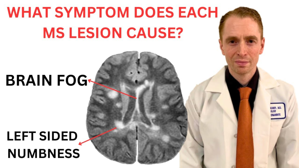
What Symptom Does Each Multiple Sclerosis MRI Lesion Cause? Neurologist Explains

My patients often ask me what symptoms would be expected based on an MS MRI lesion in a particular location. This reviews MRI scans showing common MS lesions and their associated symptoms. Keep in mind that actual symptoms vary considerably based on the amount of injury to the underlying nerve fibers associated with that lesion.

https://youtube.com/watch?v=06FzdkUywY0%3Fsi%3DJq3-V67R1cp-6qqa

Multiple Sclerosis Newsflash – Ageing reduces the ability of regulatory T cells to enhance myelin regeneration, study finds

The loss of myelin due to age or neurodegenerative diseases has profound consequences for neurological functions. At the Institute for Neurosciences UMH-CSIC, several researchers investigate the mechanisms that can help recover myelin.

OPCs (green) and myelin proteins expressed by OPCs converted into oligodendrocytes (red and white). CREDIT Instituto de Neurociencias UMH-CSIC

OPCs exposed to young regulatory T cells
IMAGE: OPCS (GREEN) AND MYELIN PROTEINS EXPRESSED BY OPCS CONVERTED INTO OLIGODENDROCYTES (RED AND WHITE).view more CREDIT: INSTITUTO DE NEUROCIENCIAS UMH-CSIC

Regulatory T lymphocytes regulate the immune system and have regenerative functions in many contexts, including myelin restoration. To determine if the function of these cells is impaired with age, Alerie Guzmán de la Fuente, a Miguel Servet investigator at the Institute for Health and Biomedical Research of Alicante (ISABIAL) and the Institute for Neurosciences (IN), a joint centre of the Miguel Hernández University (UMH) of Elche and the Spanish National Research Council (CSIC), has co-led a study, together with the researcher Denise Fitzgerald from Queen’s University Belfast (UK), demonstrating that, although regulatory T lymphocyte number increases with age, their ability to promote oligodendrocyte progenitor stem cells (OPCs) to form new cells to replace lost myelin is reduced.

Myelin is a protective layer present in the nervous system that surrounds nerve fibres, allowing quick and appropriate communication between neurons: “It is similar to the plastic that covers the copper in a cable,” explains Guzmán de la Fuente, and she points out that myelin loss linked to ageing or neurodegenerative diseases, such as multiple sclerosis, has serious consequences for neurological functions. In this work, published in the journal Nature Communications, the researchers have focused their study on how ageing, a key risk factor that limits myelin regeneration, affects the regulatory T cells regenerative functions in the brain and spinal cord.

To carry out this study, the researchers used mice between 19 and 23 months old as an animal model, which resembles an approximate age of 65 to 70 in humans. The experts detected that the presence of regulatory T lymphocytes increases with age; however, these had lost the ability to enhance the conversion of OPCs to new oligodendrocytes that regenerate myelin upon damage.

Reversible in a young environment

The researchers wanted to determine whether this loss in regulatory T cell function was completely irreversible. To do so, they carried out several experiments in young mice in which they replaced their young regulatory T lymphocytes with aged ones and verified that both young and aged cells have the same capacity to enhance myelin regeneration in a young animal.

The results of these experiments, in which researchers from the Institute for Neurosciences and ISABIAL Francisco Javier Rodríguez Baena and Sonia Cabeza Fernández, together with a team of researchers from the University of Cambridge (UK), Altos Laboratories (UK) and the University of Syddansk (Denmark), have also participated, are very positive because they suggest that the loss of function may be reversible.

New steps to study

“Regulatory T lymphocytes are very complex because they modulate the immune system, and in patients, it is not feasible to eliminate and exchange them for young cells”, says Alerie Guzmán de la Fuente, and she explains that this led the team to study in depth what was different between young and aged regulatory T cells. The objective was to identify some of the mechanisms involved in the failure to enhance myelin repair associated with regulatory T cell ageing to modulate them more specifically”, clarifies the researcher.

Combining a series of techniques, such as transcriptomic analysis, researchers have detected two new molecules involved in this process: Integrin alpha 2 (ITGA2) and the Melanoma Cell Adhesion Molecule (MCAM). They verified that both molecules not only decrease with ageing in regulatory T lymphocytes but are also involved in the interaction between these lymphocytes and the stem cells that regenerate myelin, the OPCs.

This finding opens new avenues of research to determine whether ITGA2 and MCAM molecules have potential as therapeutic targets. Future studies are needed to determine whether manipulation of these proteins can increase myelin regeneration in aged multiple sclerosis patients.

Cellular architecture of lesions in Multiple Sclerosis now mapped out

Tie One on for Multiple Sclerosis

Using advanced methodology, scientists in Sweden could reveal at the cellular level how lesions in multiple sclerosis develop. The new results are presented in the journal Cell by researchers from Karolinska Institutet and Stockholm University.

Over 1.8 million people worldwide are diagnosed with multiple sclerosis (MS). In this disease, the body’s immune cells attack the cells that form myelin, the so-called oligodendrocytes, which belong to the group of glial cells. Without myelin, signals between nerve cells cannot travel as fast as usual, resulting in symptoms such as reduced sensation and lack of coordination. MS is characterized by lesions in the brain and spinal cord.

“We wanted to understand which cells are part of the lesions and their dynamics over time,” says Petra Kukanja, co-first author in the study and PhD student at the research group of Professor Gonçalo Castelo-Branco at the Department of Medical Biochemistry and Biophysics Karolinska Institutet.

The researchers used a technology called in situ sequencing, developed in the research group of Professor Mats Nilsson at Stockholm University. This involves analyzing and identifying cells in a tissue section by reading which genes are active in a particular cell. This pattern reveals both how different cell types are arranged in the tissue, in this case, the spinal cord, and how the cells interact with each other. To study how the lesions develop, samples were taken at different times from mice experimentally induced with MS-like symptoms and from human MS patients.

“We analyzed simultaneously 239 genes and saw that active lesions in mice were built up centrifugally in two dimensions, with immune cells in the middle and different types of glial cells around them,” says Christoffer Mattsson Langseth, also co-first author of the study, and PhD student at the Department of Biochemistry and Biophysics, Stockholm University at Professor Mats Nilsson’s group.

 In the mice, it was possible to see that the lesions first appeared in the spinal cord and then spread towards the brain. In spinal cord samples from four deceased MS patients, 260 genes were simultaneously analyzed, and the cellular architecture of the lesions could be determined. The authors also found new lesions and new sub-structures within the lesions.

Previously, oligodendrocytes have mainly been seen as victims of immune cell attacks.

“The fact that they are active in the outer regions of lesions but also in the entire brain and spinal cord raises the question of whether they dampen the disease or drive it,” says Petra Kukanja.

In the next step, the researchers want to use the same methodology to analyze samples from more MS patients, as the disease is very heterogeneous. Another question is what the lesions look like when patients receive different treatments.

The two researchers highlight the extensive and fruitful collaboration between their research groups, which has been ongoing since 2019 and to which they have contributed their respective expertise – from Karolinska Institutet on the cell biology behind MS and from Stockholm University with their pioneering technology in situ sequencing and new methodology for complex image analysis.

Frexalimab for Multiple Sclerosis Explained by Neurologist

Sanofi is developing Frexalimab to treat relapsing and progressive multiple sclerosis. It targets CD40L which stimulates the proliferation of t-cells. However effective is it, and is there a risk of infection and clotting? This video reviews the result of a recent phase II trial.