
Actor and advocate Selma Blair tells Meet the Press Moderator Kristen Welker that she lives “in pain every day” after her multiple sclerosis went into remission and opens up about her treatment.
Actor and advocate Selma Blair tells Meet the Press Moderator Kristen Welker that she lives “in pain every day” after her multiple sclerosis went into remission and opens up about her treatment.
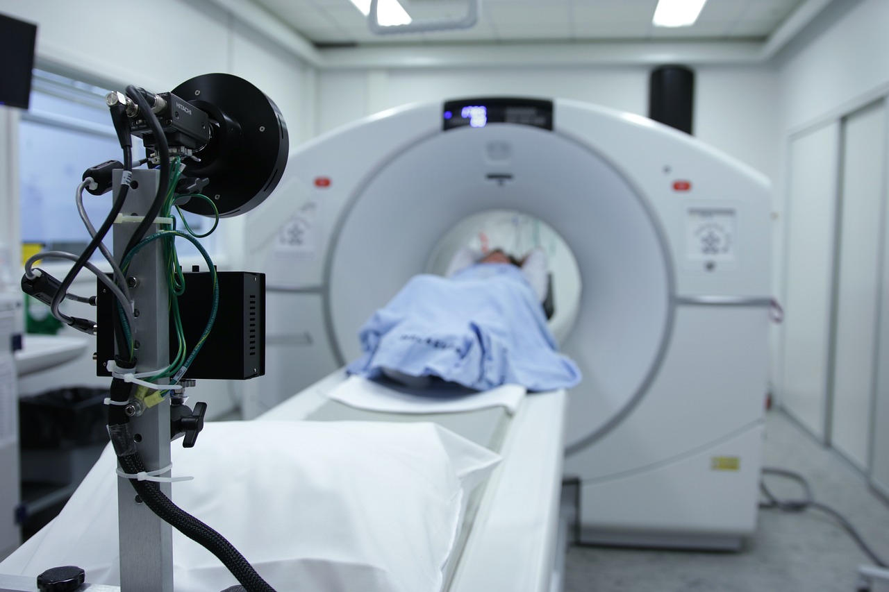
A new study from Brigham and Women’s Hospital, a founding member of the Mass General Brigham healthcare system, suggests that positron emission tomography (PET) brain scans could reveal hidden inflammation in patients with multiple sclerosis (MS) who are being treated with highly effective treatments. The findings were published in Clinical Nuclear Medicine.
“One of the perplexing challenges for clinicians treating patients with MS is after a certain amount of time, patients continue to get worse while their MRIs don’t change,” said lead author Tarun Singhal, MD, MBBS, an associate professor of Neurology in the Brigham’s Department of Neurology and director of the PET Imaging Program in the Ann Romney Center for Neurologic Diseases. “This is a new approach that is potentially going to be very helpful for the field, for research, and hopefully for clinical use.”
Singhal collaborated with others in the Brigham Multiple Sclerosis Center and the Ann Romney Center. The study started when Singhal noticed that patients treated with the most effective MS treatments were experiencing worsening symptoms. The team has worked for the past eight years on developing an approach of imaging cells called microglia. Microglia are immune cells in the brain that are thought to have a role in MS disease progression but cannot be seen by a routine MRI. The team developed a technique called F18 PBR 06 PET imaging. It involves the injection of a tracer, or dye, that binds to the microglia cells.
Rohit Bakshi, MD, of the Department of Neurology and a co-author on the paper, said increased microglial activity means more atrophy of grey matter in the brain.
“This can affect cognition, movement, fine motor skills, and other aspects of their life,” Bakshi said.
In their paper, the authors describe the term “smouldering” inflammation. Just as a smouldering fire can burn slowly without smoke or flame, smouldering inflammation may linger in patients with MS, driving disease progression and symptoms, even when it cannot be assessed on MRI.
The newly published study involved performing PET scans on 22 people with MS and eight healthy controls. Researchers measured the glial activity load on the PET scans, a new measure developed in Singhal’s lab where lab members looked at the level of smouldering inflammation from microglia in MS patients. They compared those scans to patients’ disability and fatigue levels and not only found that PET scans could show hidden inflammation caused by microglia, but the damage to patients’ brains correlated with the disability and fatigue levels they were experiencing. The researchers were also able to better classify patients with MS between high-efficacy and low-efficacy treatments. Those with low-efficacy treatments had more abnormalities on their PET scans, suggesting more microglial cell activation. Those using high-efficacy treatments had a lower degree of PET abnormality than those on no or low-efficacy treatments but still had an abnormal increase of microglial activation compared to healthy people, suggesting that while high-efficacy treatments helped to reduce neuroinflammation, there was residual inflammation despite treatment, which could account for future worsening and progression independent of relapse activity (PIRA) in these MS patients.
“Our therapies are excellent in that we’ve improved MS patients’ lives,” Bakshi said. “There’s no doubt about that, but we’re still not at the finish line.”
One limitation to the study is the initial group was small. The authors note that PET scans can also be expensive and expose patients to some level of radiation, whereas MRIs do not. Singhal said that radiation could be reduced because of the long half-life and the requirement for a lower administered dose of the F18 PBR06 tracer. The tracer also produces better imaging characteristics than previously used tracers with shorter half-lives.
Bakshi said despite the limitations, the study shines an important light on the power of PET scanning, specifically to find microglial activation.
“This study tells us something new about the disease and may be giving us an important clue as to what is driving disease progression in patients,” he said.
Singhal said before the technique can be used routinely in a clinical setting, it must be validated on a larger sample size. Other longer half-life PET tracers have been approved by the FDA for clinical use, for example, amyloid PET tracers for studying Alzheimer’s disease. If approved, [F-18]PBR06 could also be used to personalize and predict a patient’s treatment course in MS and other brain diseases. However, the authors note that even before approval, [F-18]PBR06 can help advance drug development and perform multicentric clinical trials.
“It’s very exciting that our novel approach worked and correlated so strongly with clinical measures we assessed,” he said. “It means our approach is relevant clinically.”
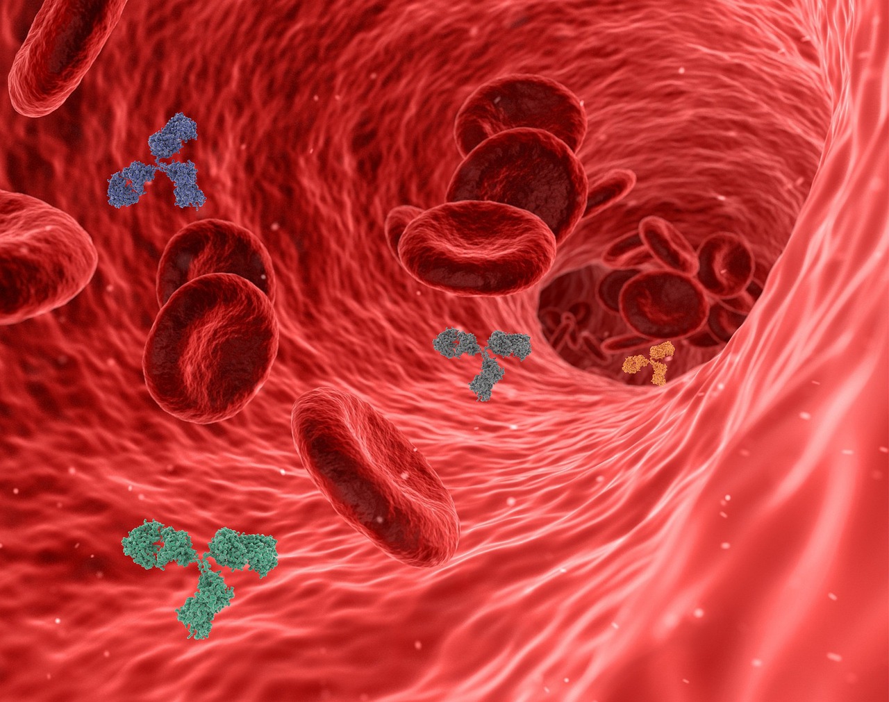
Autoantibodies are antibodies that are supposed to fight off invaders but end up turning against one’s own body, causing problems like autoimmune diseases. Utilizing the U.S. Department of Defense Serum Repository, a cohort encompassing more than 10 million individuals, researchers conducted whole-proteome autoantibody profiling on hundreds of MS patients’ samples collected before and after symptom onset. They discovered a distinct cluster of patients exhibiting an autoantibody signature targeting a common recognizable pattern. Notably, these patients showed antibody reactivity years before developing any MS symptoms and had elevated levels of serum neurofilament light (sNfL), indicating early neuroaxonal injury.
Danillo Augusto, Ph.D., an assistant professor in biology at the University of North Carolina at Charlotte and a co-author of the study, stated, “This study sheds light on the preclinical phase of MS and provides a promising avenue for early detection and intervention. Identifying patients at high risk of developing MS before symptom onset could revolutionize patient care and treatment strategies.”
Further validation of this autoantibody signature was conducted on samples from a separate MS cohort, confirming its high specificity for patients diagnosed with MS. This finding marks a significant milestone in MS research, potentially paving the way for the development of antigen-specific biomarkers for high-risk individuals with clinically or radiologically isolated neuroinflammatory syndromes.
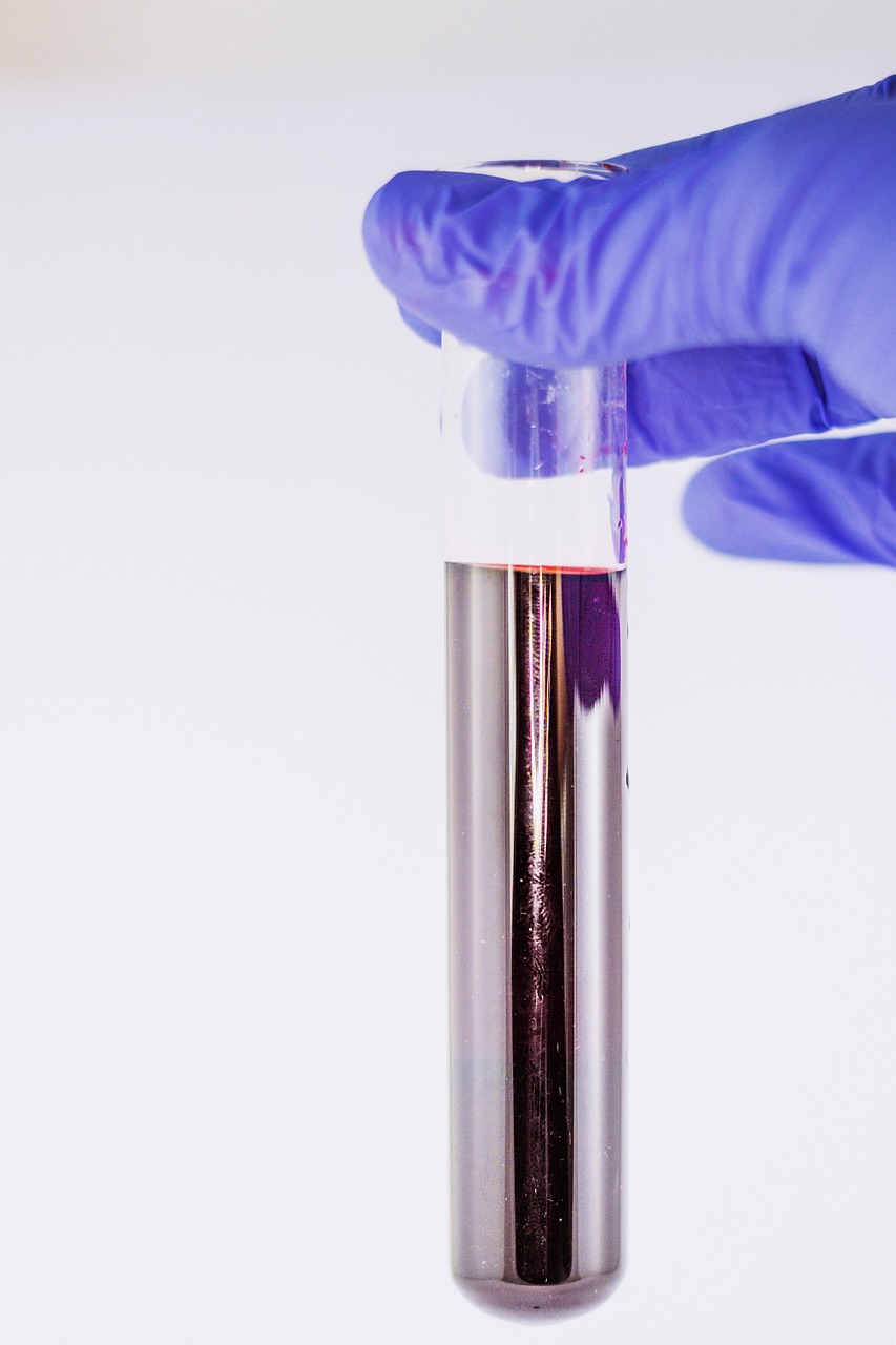
In a discovery that could hasten treatment for patients with multiple sclerosis (MS), UC San Francisco scientists have discovered a harbinger in the blood of some people who later went on to develop the disease.
In about 1 in 10 cases of MS, the body begins producing a distinctive set of antibodies against its own proteins years before symptoms emerge. These autoantibodies appear to bind to both human cells and common pathogens, possibly explaining the immune attacks on the brain and spinal cord that are the hallmark of MS.
The findings were published in Nature Medicine on April 19.
MS can lead to a devastating loss of motor control, although new treatments can slow the progress of the disease and, for example, preserve a patient’s ability to walk. The scientists hope the autoantibodies they have discovered will one day be detected with a simple blood test, giving patients a head start on receiving treatment.
“Over the last few decades, there’s been a move in the field to treat MS earlier and more aggressively with newer, more potent therapies,” said UCSF neurologist Michael Wilson, MD, a senior author of the paper. “A diagnostic result like this makes such early intervention more likely, giving patients hope for a better life.”
Linking infections with autoimmune disease
Autoimmune diseases like MS are believed to result, in part, from rare immune reactions to common infections.
In 2014, Wilson joined forces with Joe DeRisi, PhD, president of the Chan Zuckerberg Biohub SF and a senior author of the paper, to develop better tools for unmasking the culprits behind autoimmune disease. They took a technique in which viruses are engineered to display bits of proteins like flags on their surface, called phage display immunoprecipitation sequencing (PhIP-Seq), and further optimized it to screen human blood for autoantibodies.
PhIP-Seq detects autoantibodies against more than 10,000 human proteins, enough to investigate nearly any autoimmune disease. In 2019, they successfully used it to discover a rare autoimmune disease that seemed to arise from testicular cancer.
MS affects more than 900,000 people in the US. Its early symptoms, like dizziness, spasms, and fatigue, can resemble other conditions, and diagnosis requires careful analysis of brain MRI scans.
The phage display system, the scientists reasoned, could reveal the autoantibodies behind the immune attacks of MS and create new opportunities to understand and treat the disease.
The project was spearheaded by first co-authors Colin Zamecnik, PhD, a postdoctoral researcher in DeRisi’s and Wilson’s labs; and Gavin Sowa, MD, MS, former UCSF medical student and now internal medicine resident at Northwestern University.
They partnered with Mitch Wallin, MD, MPH, from the University of Maryland and a senior author of the paper, to search for autoantibodies in the blood of people with MS. These samples were obtained from the U.S. Department of Defense Serum Repository, which stores blood taken from armed service members when they apply to join the military.
The group analyzed blood from 250 MS patients collected after their diagnosis, plus samples taken five or more years earlier when they joined the military. The researchers also looked at comparable blood samples from 250 healthy veterans.
Between the large number of subjects and the before-and-after timing of the samples, it was “a phenomenal cohort of individuals to look at to see how this kind of autoimmunity develops over the course of clinical onset of this disease,” said Zamecnik.
A consistent signature of MS
Using a mere one-thousandth of a milliliter of blood from each time point, the scientists thought they would see a jump in autoantibodies as the first symptoms of MS appeared.
Instead, they found that 10% of the MS patients had a striking abundance of autoantibodies years before their diagnosis.
The dozen or so autoantibodies all stuck to a chemical pattern that resembled one found in common viruses, including Epstein-Barr Virus (EBV), which infects more than 85% of all people, yet has been flagged in previous studies as a contributing cause for MS.
Years before diagnosis, this subset of MS patients had other signs of an immune war in the brain. Ahmed Abdelhak, MD, co-author of the paper and a postdoctoral researcher in the UCSF laboratory of Ari Green, MD, found that patients with these autoantibodies had elevated levels of neurofilament light (Nfl), a protein that gets released as neurons break down.
Perhaps, the researchers speculated, the immune system was mistaking friendly human proteins for some viral foe, leading to a lifetime of MS.
“When we analyze healthy people using our technology, everybody looks unique, with their own fingerprint of immunological experience, like a snowflake,” DeRisi said. “It’s when the immunological signature of a person looks like someone else, and they stop looking like snowflakes that we begin to suspect something is wrong, and that’s what we found in these MS patients.”
A test to speed patients toward the right therapies
To confirm their findings, the team analyzed blood samples from patients in the UCSF ORIGINS study. These patients all had neurological symptoms and many, but not all, went on to be diagnosed with MS.
Once again, 10% of the patients in the ORIGINS study who were diagnosed with MS had the same autoantibody pattern. The pattern was 100% predictive of an MS diagnosis. Across both the Department of Defense group and the ORIGINS group, every patient with this autoantibody pattern had MS.
“Diagnosis is not always straightforward for MS, because we haven’t had disease specific biomarkers,” Wilson said. “We’re excited to have anything that can give more diagnostic certainty earlier on, to have a concrete discussion about whether to start treatment for each patient.”
Many questions remain about MS, ranging from what’s instigating the immune response in some MS patients to how the disease develops in the other 90% of patients. But the researchers believe they now have a definitive sign that MS is brewing.
“Imagine if we could diagnose MS before some patients reach the clinic,” said Stephen Hauser, MD, director of the UCSF Weill Institute for Neurosciences and a senior author of the paper. “It enhances our chances of moving from suppression to cure.”