All in the Eyes: High-Resolution Retinal Maps Aid in Diagnosing Diseases – A Beacon of Hope for Those with Multiple Sclerosis or Diabetes

Retinal Maps

Multiple retinal maps of single eyes with white indicate where the retina is thickest, and coloured areas represent thinner regions of the retina. The research identified 294 genes influencing the contours of these retinal maps and pinpointed specific areas of the maps linked to disease. Credit WEHI

For individuals managing conditions like multiple sclerosis or diabetes, early detection and effective management are crucial. Excitingly, a groundbreaking study led by the Walter and Eliza Hall Institute (WEHI) has revealed how high-resolution retinal maps can play a vital role in diagnosing these and other diseases.

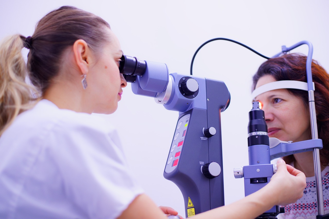
Researchers conducted one of the largest eye studies ever, analyzing over 50,000 eyes using advanced artificial intelligence technology. This innovative approach produced incredibly detailed maps of the retina, revealing how variations in retinal thickness are linked to a range of diseases, including type 2 diabetes, dementia, and multiple sclerosis.

Key Findings and Potential Benefits

The study’s results open up new possibilities for routine eyecare imaging as a powerful tool for disease screening and management. Much like mammograms have become essential for breast cancer detection, these retinal maps could serve as non-invasive diagnostic biomarkers for numerous conditions.

Here’s a quick overview of the groundbreaking findings:

  • AI-powered research has created the most detailed retinal maps ever produced.
  • These maps reveal critical connections between retinal thinning and various diseases.
  • New genetic factors influencing retinal thickness have been identified.

The Retina: A Window to the Brain

The retina is part of the central nervous system, including the brain and spinal cord. Many diseases, particularly neurological and metabolic disorders like multiple sclerosis and diabetes, are linked to the degeneration or disruption of this system. With over 3 billion people worldwide living with a brain-related condition, this research holds immense significance.

Lead researcher Dr. Vicki Jackson from WEHI emphasized the importance of retinal imaging: “We’ve shown that retinal imaging can act as a window to the brain, detecting associations with neurological disorders like multiple sclerosis and many other conditions.”

Unveiling New Genetic Insights

The study also identified 294 genes that influence retinal thickness, shedding light on their role in the development and growth of the retina. These genetic factors could provide valuable insights into the progression of diseases and aid in early detection.

Pioneering the Future of Diagnostic Tools

Professor Melanie Bahlo, the study lead and bioinformatician at WEHI, highlighted the transformative power of AI: “Technologies like AI fuel discovery, and when fused with brilliant minds, there is an extraordinary ability to transform big population data into far-reaching insights.”

This research reinforces the growing field of oculomics, which uses the eye to diagnose health conditions. By harnessing the power of AI and big data, scientists are paving the way for innovative, non-invasive approaches to predicting and diagnosing diseases.

The study involved numerous collaborators, including the UK Biobank, University of Washington, Lowy Medical Research Institute, Moorfields Eye Hospital, and University College London.

The findings underscore the potential for high-resolution retinal maps to revolutionize disease diagnosis and management, offering new hope for individuals with multiple sclerosis, diabetes, and other conditions. As researchers continue to explore the complex links between retinal health and systemic diseases, the future of medical diagnostics looks promising.

Breakthrough Drug Could Restore Vision for Multiple Sclerosis Patients!

Researchers funded to examine how different genes associated with autism spectrum disorders may similarly impact our brain’s neurons, resulting in heightened sensitivity to sounds.

New Hope in MS Treatment from University of Colorado Anschutz Medical Campus

Researchers have developed a promising new drug that could restore vision for people suffering from multiple sclerosis (MS) and other neurological conditions. This breakthrough, published in the journal Nature Communications, involves a drug called LL-341070.

How It Works:

The drug boosts the brain’s ability to repair damaged myelin—the protective sheath around nerve fibers. Myelin damage is a key factor in diseases like MS, leading to vision loss, motor skill impairment, and cognitive decline.

Key Findings:

  • Enhanced Brain Repair: LL-341070 significantly sped up the repair process, improving vision-related brain function in mice.
  • Potential for MS Patients: By accelerating natural repair mechanisms, the drug offers hope for reversing some of the damage caused by MS.
  • Partial repair is impactful: Even partial myelin repair led to significant improvement in vision-related brain functions.

Researchers’ Insight:

“This research brings us closer to a world where the brain has the capacity to heal itself,” says Ethan Hughes, PhD. “By harnessing this potential, we hope to help people with diseases like MS regain their vision and cognitive function.”

Next Steps:

The researchers plan to test LL-341070 in other areas of the brain and refine the treatment to make it more effective and accessible to patients.

The Future:

The discovery of LL-341070 is just the beginning. As Dr. Hughes puts it, “We are optimistic that LL-341070 and similar therapies could one day provide real, tangible benefits to patients by improving overall brain function and quality of life.”

This could be a game-changer for people with MS, offering a new ray of hope for restoring lost vision.

Study finds drug may slow progression of diabetic retinopathy in diabetics

Empagliflozin was associated with a 22 percent lower risk of progression from early stages of diabetic retinopathy to more advanced stages in this patient group
Empagliflozin was associated with a 22 per cent lower risk of progression from early stages of diabetic retinopathy to more advanced stages in this patient group.


Diabetic retinopathy (DR) affects about 26 per cent of people with diabetes. While many patients have an early form of DR, the condition can progress, posing the threat of irreversible vision loss. Empagliflozin is a sodium-glucose contratransporter-2 inhibitor (SGLT-2), an oral medication that helps patients with type 2 diabetes control their glucose levels. A research team led by investigators from Mass General Brigham conducted a cohort study comparing treatment with empagliflozin to another form of diabetes treatment for patients with type 2 diabetes. They found that while empagliflozin did not reduce the risk of DR in patients with no history of that disease, among patients who had early-stage DR, the medication was associated with a decreased risk of progression.


“Our findings showed that in patients with type 2 diabetes and non-proliferative diabetic retinopathy, empagliflozin could be beneficial in slowing down progression to more advanced stages of diabetic retinopathy,” said lead author Helen Tesfaye, PharmD, MSc, of the Division of Pharmacoepidemiology at Brigham and Women’s Hospital. “Our findings could help inform clinical decision-making for patients with diabetes who have non-proliferative diabetic retinopathy or are at risk for developing DR.”

The study represents a collaboration between Brigham and Women’s Hospital (BWH) and Massachusetts General Hospital (MGH), both founding members of the Mass General Brigham healthcare system, with experts from BWH’s Division of Renal (Kidney) Medicine and MGH’s Diabetes Center working with Tesfaye and colleagues from the Division of Pharmacoepidemiology.

“Leveraging the clinical and analytical expertise of our multidisciplinary team allows us to identify risks and benefits of diabetes medications that are not being studied—or may not be apparent—in clinical trials,” said Deborah Wexler.

A new study reveals a breakthrough in the treatment of visual and cognitive impairments in multiple sclerosis.

A pilot study conducted by researchers at the Kessler Foundation showed promising results in using methylphenidate to improve oculomotor function in individuals with multiple sclerosis (MS). The findings could have significant implications for the treatment of visual and cognitive impairments associated with multiple sclerosis (MS).

Silvana Costa, PhD

Dr. Costa is a research scientist at the Centers for Neuropsychology, Neuroscience, and Multiple Sclerosis Research at the Kessler Foundation. CREDIT Kessler Foundation

In a pilot randomized placebo-controlled trial, 11 participants with MS were randomly assigned to receive either methylphenidate or a placebo for four weeks. After a seven-day washout period, they underwent a crossover treatment for four more weeks. Assessments of oculomotor speed using the King-Devick test and information processing speed were conducted before and after each treatment phase.

“We noticed a significant increase in the participants’ eye movement speed when they were given methylphenidate compared to when they were given the placebo,” said Timothy J. Rich, PhD, OTR/L, research scientist at the Center for Stroke Rehabilitation Research at Kessler Foundation. “This improvement in eye movement speed was directly linked to better performance on tasks that involve hand-eye coordination, which is important for everyday activities.”

Dr. Silvana L Costa, who works as a research scientist at the Centers for Neuropsychology, Neuroscience, and Multiple Sclerosis Research at Kessler Foundation, and is also a co-author of the study, stated, “Our results indicate that methylphenidate might offer valuable benefits as a treatment for individuals with MS, particularly for those who are dealing with oculomotor deficits impacting their visual processing speed and, consequently, their overall quality of life.”

“These results are promising,” noted Dr. Costa, “offering a potential pathway to mitigate some of the cognitive and visual symptoms that are prevalent in MS.”

How does fibromyalgia affect the eyes?

Fibromyalgia is a chronic disease that causes severely disabling muscle pain. The nature of this pain is not known, and it is accompanied by other symptoms: severe fatigue, memory and concentration problems, sleep disorders, anxiety, etc. Other symptoms, such as problems in the eye, which are often not discussed, may also develop. But what is the link between the eyes and fibromyalgia? What are the different eye disorders the disease can cause? And what can be done about it?