Keratosis pilaris – the signs, symptoms and treatments of this condition

Keratosis pilaris or chicken skin

Keratosis pilaris or chicken sk

Keratosis pilaris is a common and harmless condition where the skin becomes rough and bumpy, as if covered in permanent goose pimples.

There’s no cure for keratosis pilaris, but you may be able to improve the rash by using soap-free cleansers, moisturising, and gently removing dead skin cells from the surface of the skin (exfoliating).

There’s no real need to see your GP unless the condition is causing you concern. It usually improves as you get older and sometimes disappears completely in adulthood.

This page provides more information on keratosis pilaris and explains what you can do if you have the condition.

Where keratosis pilaris occurs

Keratosis pilaris most commonly affects the back of the upper arms, and sometimes the buttocks and the front of the thighs. Less often, the forearms and upper back may be affected.

There are also rare variants of keratosis pilaris that can affect the eyebrows, face and scalp, or the entire body.

How it affects the skin

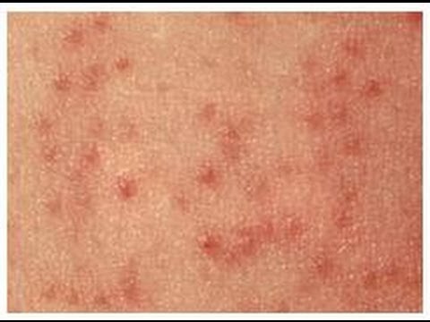
The patches of affected skin will be covered in tiny spiky bumps, which may be white, red or skin-coloured. This spotting looks like “chicken skin” or permanent goose pimples, and the skin feels rough, like sandpaper.

In some people, the skin itches and there may be inflammation and pinkness around the bumps.

Keratosis pilaris can’t be spread from person to person (contagious).

The skin tends to improve in summer and get worse during winter months or dry conditions.

Who’s affected

Keratosis pilaris is very common, affecting up to one in three people in the UK.

It can affect people of all ages, but it’s particularly common in:

children and adolescents

females

people with eczema or a condition called ichthyosis

people of Celtic origin

The condition typically starts during childhood, although it can sometimes occur in babies, and gets worse in adolescence, around puberty.

Keratosis pilaris sometimes improves after puberty, and may even disappear in adulthood, although many adults still have the condition in their 40s and 50s. It’s uncommon in elderly people.

What causes keratosis pilaris?

Keratosis pilaris runs in families and is inherited from your parents. If one parent has the condition, there’s a one in two chance that any children they have will also inherit it.

Keratosis pilaris occurs when too much keratin builds up in the skin’s hair follicles. Keratin is a protein found in the tough outer layer of skin, which causes the surface of the skin to thicken, hence the name “keratosis”.

The excess keratin blocks the hair follicles with plugs of hard, rough skin. The tiny plugs widen the pores, giving the skin a spotty appearance.

It’s often associated with other dry skin conditions, such as eczema and ichthyosis, which make the rash worse.

Treating keratosis pilaris

There’s little that can be done to treat keratosis pilaris, and it often gets better on its own without treatment.

However, if it’s bothering you, the following measures may help improve your rash:

use non-soap cleansers rather than soap – ordinary soap may dry your skin out and make the condition worse

moisturise your skin when it’s dry – your GP or pharmacist can recommend a suitable cream, although moisturisers and emollients only reduce the dryness of your skin and won’t cure the rash; creams containing salicylic acid, lactic acid or urea are thought to be the most effective

gently rub the skin with an exfoliating foam pad or pumice stone to exfoliate the rough skin – be careful not to scrub too hard and rub off layers of skin

take lukewarm showers rather than hot baths

You can also ask your GP about “off-label” treatments that may help. These are products that haven’t been officially approved and licensed for treating keratosis pilaris, but are sometimes used because they’ve helped people in the past.

Examples include:

creams containing retinol, which is derived from vitamin A

chemical peels

microdermabrasion – a cosmetic exfoliation treatment that is sometimes offered in health spas

There’s no strong evidence to suggest that these therapies are effective treatments for keratosis pilaris. You may also have to pay for some of them privately.

Kyphosis – an introduction

Kyphosis

Kyphosis

Kyphosis is curvature of the spine that causes the top of the back to appear more rounded than normal.

Everyone has some degree of curvature in their spine. However, a curve of more than 45 degrees is considered excessive.

Sometimes kyphosis doesn’t cause any symptoms other than the back appearing abnormally curved or hunched. However, in some cases the condition causes:

back pain and stiffness

tenderness of the spine

tiredness

Back pain can be particularly problematic in adults with kyphosis because the body has to compensate for the spinal abnormality.

If you have severe kyphosis, your symptoms may get worse over time. You may also have difficulty breathing and eating.

What causes kyphosis?

In kyphosis, the normal curve in the middle section of vertebral column (the thoracic vertebrae) is more curved than normal. There are a number of reasons why this might happen, including:

poor posture (postural kyphosis) – slouching, leaning back in chairs and carrying heavy bags can stretch supporting muscles and ligaments, which can increase spinal curvature

abnormally shaped vertebrae (Scheuermann’s kyphosis) – if the vertebrae don’t develop properly, they can end up being out of position

abnormal development of the spine in the womb (congenital kyphosis) – if something disrupts the spine’s normal development, two or more vertebrae sometimes fuse together

age – as people get older, their spinal curvature can be expected to increase

Kyphosis can also develop as a result of a spinal injury.

Read more about the causes of kyphosis.

Treating kyphosis

If you have kyphosis, your treatment depends on how curved your spine is, whether you have any additional symptoms such as back pain, and the underlying causes.

Children with kyphosis may be able to be treated using non-surgical methods, such as bracing, to limit the progression of kyphosis as they grow. Treatment for mild kyphosis may not be necessary.

Kyphosis rarely requires surgical treatment. It’s only needed in some severe cases to correct the curvature of the spine.

Read more about treating kyphosis.

Emotional issues

Older children with kyphosis may become concerned or embarrassed about the effect the condition has on their appearance, or having to wear a back brace.

These concerns can affect different children in different ways. Some children can become socially withdrawn and they may be reluctant to take part in activities, such as PE, where their condition may be exposed.

There are no easy answers to these problems, but it can sometimes help to reassure your child that their feelings will improve with time.

Complications

Complications of kyphosis usually only occur in more severe cases. They include:

persistent pain that can’t be controlled with medication

breathing difficulties caused by the spine compressing the lungs and airways

Occasionally, people with kyphosis can have difficulties when the nerves running through the spine become compressed or pinched. This can disrupt nerve signals and cause symptoms such as:

numbness or weakness in the arms and legs

problems with sense of balance

loss of normal bladder or bowel control

These serious complications require urgent medical attention and surgery would usually be recommended.

Can kyphosis be prevented?

Postural kyphosis can be prevented by being aware of your posture and by taking care of your back. You should encourage your child to:

avoid slouching

sit correctly – sit upright, ensuring that the small of the back is supported

avoid carrying heavy schoolbags that can pull on the back muscles and ligaments; the best schoolbags are well-designed backpacks

take regular exercise (see below) to help strengthen the back and keep it flexible; activities such as swimming, running, walking, yoga and pilates are ideal for helping to prevent back problems

Intracranial hypertension – causes, signs and treatments

Intracranial hypertension

Intracranial hypertension

Intracranial hypertension

Intracranial hypertension (IH) is the medical name for a build-up of pressure around the brain.

It can come on suddenly – for example, as the result of a severe head injurystroke or brain abscess. This is known as acute IH.

It can also be a persistent, long-lasting problem – known as chronic IH. This is rare and sometimes it’s not clear why it happens.


Symptoms of chronic IH

Symptoms of chronic IH can include:

a constant throbbing headache – this may be worse in the morning, or when coughing or straining; it may improve when standing up

blurred or double vision

temporary loss of vision – your vision may become dark or “greyed out” for a few seconds at a time; this can be triggered by coughing, sneezing or bending down

feeling and being sick

drowsiness and irritability

Chronic IH can sometimes result in permanent vision loss, although treatment can help reduce the chances of this happening.

Causes of chronic IH

Possible causes of chronic IH include:

a blood clot on the surface of the brain – known as a chronic subdural haematoma

a brain tumour

a brain infection – such as meningitis or encephalitis

hydrocephalus – a build-up of fluid around and inside the brain

blood vessel abnormalities – such as an arteriovenous fistula or arteriovenous malformation

a blood clot in one of the veins of the brain – known as a venous sinus thrombosis

Rare causes include a blockage in the circulation of fluid at the bottom of the skull (Chiari malformation), inflammation of the blood vessels in the brain (vasculitis) and abnormal skull growth in children (craniosynostosis).

Idiopathic IH

In many cases, the cause of chronic IH is unclear. This is known as idiopathic IH, or sometimes benign IH.

It mainly affects women in their 20s and 30s, and has been associated with:

being overweight or obese – most cases occur in overweight women, although it’s not clear why

hormone problems – such as Cushing’s syndrome, hypoparathyroidism, an underactive thyroid or an overactive thyroid

certain medications – including some antibiotics, steroid medication and contraceptive pills

a lack of red blood cells (iron deficiency anaemia) or too many red blood cells (polycythaemia vera)

chronic kidney disease

lupus – a problem with the immune system

But these are only linked with idiopathic IH, they’re not necessarily causes. You can find a full list of conditions and medications associated with idiopathic IH (PDF, 21kb) on the IIH UK website.

Tests for chronic IH

IH may be suspected if you have symptoms of increased pressure on your brain, such as vision problems and headaches.

Several tests may be carried out to diagnose it, such as:

an examination to check functions like your muscle strength, reflexes and balance – any issues could be a sign of a problem with your brain or nerves

an assessment of your eyes and vision

a computerised tomography (CT) scan or magnetic resonance imaging (MRI) scan of your brain

a lumbar puncture – where a needle is inserted into your spine – to check for high pressure in the fluid that surrounds your brain and spinal cord

Idiopathic IH may be diagnosed if you have increased pressure on your brain and no other cause can be found.

Treatments for chronic IH

Treatment for IH depends on the underlying cause, if this is known.

The main treatments for idiopathic IH are:

losing weight if you’re overweight – this can often help reduce symptoms and may sometimes relieve them altogether

stopping any medication that may be causing your symptoms

medication to remove excess fluid from the body (diuretics)

medication to reduce the production of cerebrospinal fluid in your brain

a short course of steroid medication to relieve headaches and reduce the risk of vision loss

regular lumbar punctures to remove excess fluid from your spine and help reduce the pressure on your brain

Surgery

Surgery may be considered if other treatments don’t help.

The main types of surgery for chronic IH are:

shunt surgery – a thin, flexible tube is inserted into the fluid-filled space in your skull or spine to divert excess fluid to another part of your body

optic nerve sheath fenestration – the protective layer surrounding your optic nerve, the nerve connecting the eye to the brain, is opened up to relieve pressure on it and allow fluid to drain away

These procedures can provide relief from your symptoms, but they also carry a risk of potentially serious complications. Talk to your surgeon about what your operation involves and what the risks are.

Outlook for chronic IH

Chronic IH can be life threatening if it remains undiagnosed and the underlying causes are not treated. You should be referred to a specialist as soon as possible if your doctor suspects it.

Idiopathic IH isn’t usually life threatening, but can be a lifelong problem. While many people find their symptoms are relieved with treatment, the symptoms can recur and can have a significant impact on your life.

There’s also a risk that you could lose your vision, even though treatment can help reduce this risk.

Permanent vision loss is estimated to occur in between 1 in every 5 to 20 people with idiopathic IH.

Hemochromatosis – the signs and symptoms

Hemochromatosis

Hemochromatosis

Symptoms

Symptoms of haemochromatosis usually begin between the ages of 30 and 60, although they can occasionally occur earlier.

The symptoms tend to develop earlier in men than in women. Women often don’t experience problems until after the menopause.

Sometimes there are no symptoms and the condition is only found during a blood test.

Early symptoms

Initial symptoms of haemochromatosis can include:

feeling very tired all the time (fatigue)

weight loss

weakness

joint pain

in men, an inability to get or maintain an erection (erectile dysfunction)

in women, irregular periods or absent periods

These symptoms can have many different causes and may sometimes just be put down to getting older.

Later problems

As the condition progresses, it can also cause problems such as:

loss of sex drive (libido)

darkening of the skin – you may look permanently tanned

tummy (abdominal) pain and swelling

yellowing of the skin and eyes (jaundice)

feeling thirsty all the time and needing to pee frequently

severe pain and stiffness in your joints, particularly in the fingers

chest pain

shortness of breath

swelling of your hands and feet

an irregular heartbeat (arrhythmia)

in men, the testicles getting smaller

These problems are often caused by complications of haemochromatosis that can occur if the condition isn’t treated early on.

When to see your GP

See your GP if you have:

persistent or worrying symptoms that could be caused by haemochromatosis – particularly if you have a northern European family background, as the condition is most common in this group

a parent or sibling with haemochromatosis, even if you don’t have symptoms yourself – tests can be done to check if you’re at risk of developing problems

Talk to your GP about whether you should have blood tests to check for haemochromatosis.

Read more about how haemochromatosis is diagnosed.

Gout – what are the signs and symptoms of gout

Signs and Symptoms of Gout

Signs and Symptoms of Gout

Symptoms of gout

The main symptom of gout is a sudden attack of severe pain in one or more joints, typically your big toe.

Other symptoms can include:

the joint feeling hot and very tender, to the point of being unable to bear anything touching it
swelling in and around the affected joint
red, shiny skin over the affected joint
peeling, itchy and flaky skin as the swelling goes down


The intense pain can make getting around difficult. Even the light pressure of a bed cover or blanket can be unbearable.

Which joints can be affected?

Gout can affect almost any joint and can occur in more than one joint at the same time.

The joints towards the ends of the limbs tend to be affected more often, including the:

toes – particularly the big toe joint

midfoot (where your shoelaces sit)

ankles

knees

fingers

wrists

elbows

If gout is left untreated, it’s likely to affect more joints over time.

Pattern of symptoms

Attacks of gout tend to:

occur at night, although they can happen at any time

develop quickly over a few hours

last between three and 10 days – after this time, the affected joint should start to return to normal, but the problem can persist if treatment isn’t started early

come back – you may experience attacks every few months or years

become more frequent over time if not treated

It’s difficult to predict how often attacks will occur and when exactly they will happen.