Early-stage stem cell therapy trial shows promise for treating progressive MS

Tie One on for Multiple Sclerosis

An international team has shown that the injection of a type of stem cell into the brains of patients living with progressive multiple sclerosis (MS) is safe, well tolerated and has a long-lasting effect that appears to protect the brain from further damage.

The study, led by scientists at the University of Cambridge, University of Milan Bicocca and Hospital Casa Sollievo della Sofferenza (Italy), is a step towards developing an advanced cell therapy treatment for progressive MS.

Over 2 million people live with MS worldwide, and while treatments exist that can reduce the severity and frequency of relapses, two-thirds of MS patients still transition into a debilitating secondary progressive phase of disease within 25-30 years of diagnosis, where disability grows steadily worse.

In MS, the body’s own immune system attacks and damages myelin, the protective sheath around nerve fibres, causing disruption to messages sent around the brain and spinal cord.

Key immune cells involved in this process are macrophages (literally ‘big eaters’), which ordinarily attack and rid the body of unwanted intruders. A type of macrophage known as a microglial cell is found throughout the brain and spinal cord. In progressive forms of MS, they attack the central nervous system (CNS), causing chronic inflammation and damage to nerve cells.

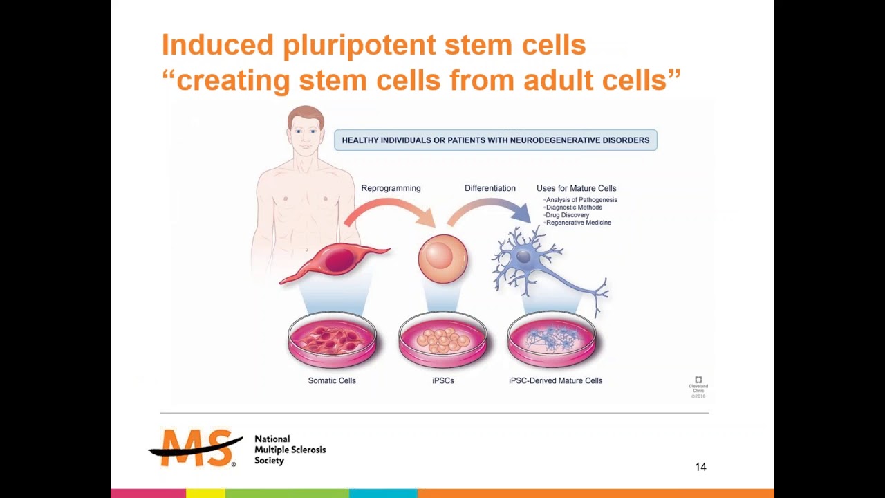
Recent advances have raised expectations that stem cell therapies might help ameliorate this damage. These involve the transplantation of stem cells, the body’s ‘master cells’, which can be programmed to develop into almost any cell type within the body.

Previous work from the Cambridge team has shown in mice that skin cells re-programmed into brain stem cells, and transplanted into the central nervous system, can help reduce inflammation and may be able to help repair damage caused by MS.

Now, in research published in the Cell Stem Cell, scientists have completed a first-in-man, early-stage clinical trial that involved injecting neural stem cells directly into the brains of 15 patients with secondary MS recruited from two hospitals in Italy. The trial was conducted by teams at the University of Cambridge, Milan Bicocca and the Hospitals Casa Sollievo della Sofferenza and S. Maria Terni  (IT) and Ente Ospedaliero Cantonale (Lugano, Switzerland) and the University of Colorado (USA).

The stem cells were derived from cells taken from brain tissue from a single, miscarried foetal donor. The Italian team had previously shown that it would be possible to produce a virtually limitless supply of these stem cells from a single donor – and in future it may be possible to derive these cells directly from the patient – helping to overcome practical problems associated with the use of allogeneic foetal tissue.

The team followed the patients over 12 months, during which time they observed no treatment-related deaths or serious adverse events. While some side-effects were observed, all were either temporary or reversible.

All the patients showed high levels of disability at the start of the trial – most required a wheelchair, for example – but during the 12 month follow up period none showed any increase in disability or a worsening of symptoms. None of the patients reported symptoms that suggested a relapse and nor did their cognitive function worsen significantly during the study. Overall, say the researchers, this points to a substantial stability of the disease, without signs of progression, though the high levels of disability at the start of the trial make this difficult to confirm.

The researchers assessed a subgroup of patients for changes in the volume of brain tissue associated with disease progression. They found that the larger the dose of injected stem cells, the smaller the reduction in this brain volume over time. They speculate that this may be because the stem cell transplant dampened inflammation.

The team also looked for signs that the stem cells were having a neuroprotective effect – protecting nerve cells from further damage. Their previous work showed how tweaking metabolism – how the body produces energy – can reprogram microglia from ‘bad’ to ‘good’. This new study looked at how the brain’s metabolism changes after the treatment. They measured changes in the fluid around the brain and in the blood over time and found certain signs linked to how the brain processes fatty acids. These signs were connected to how well the treatment works and how the disease develops. The higher the dose of stem cells, the greater the levels of fatty acids, which also persisted over the 12-month period.

Professor Stefano Pluchino from the University of Cambridge, who co-led the study, said: “We desperately need to develop new treatments for secondary progressive MS, and I am cautiously very excited about our findings, which are a step towards developing a cell therapy for treating MS.

“We recognise that our study has limitations – it was only a small study and there may have been confounding effects from the immunosuppressant drugs, for example – but the fact that our treatment was safe and that its effects lasted over the 12 months of the trial means that we can proceed to the next stage of clinical trials.”

Co-leader Professor Angelo Vescovi from the University of Milano-Bicocca said: “It has taken nearly three decades to translate the discovery of brain stem cells into this experimental therapeutic treatment This study will add to the increasing excitement in this field and pave the way to broader efficacy studies, soon to come.”

Caitlin Astbury, Research Communications Manager at the MS Society, says: “This is a really exciting study which builds on previous research funded by us. These results show that special stem cells injected into the brain were safe and well-tolerated by people with secondary progressive MS. They also suggest this treatment approach might even stabilise disability progression. We’ve known for some time that this method has the potential to help protect the brain from progression in MS.

“This was a very small, early-stage study and we need further clinical trials to find out if this treatment has a beneficial effect on the condition. But this is an encouraging step towards a new way of treating some people with MS.” 

Research advances one step closer to stem cell therapy for type 1 diabetes

This image shows functional beta cells made from human pluripotent stem cells. Insulin (red) and NKX6.1 (green) indicate two proteins produced by beta cells. CREDIT Salk Institute

 Type 1 diabetes, which arises when the pancreas doesn’t create enough insulin to control levels of glucose in the blood, is a disease that currently has no cure and is difficult for most patients to manage. Scientists at the Salk Institute are developing a promising approach for treating it: using stem cells to create insulin-producing cells (called beta cells) that could replace nonfunctional pancreatic cells.

In a study published on June 7, 2021, in the journal Nature Communications, the investigators reported that they have developed a new way to create beta cells that is much more efficient than previous methods. Additionally, when these beta cells were tested in a mouse model of type 1 diabetes, the animals’ blood sugar was brought under control within about two weeks.

“Stem cells are an extremely promising approach for developing many cell therapies, including better treatments for type 1 diabetes,” says Salk Professor Juan Carlos Izpisua Belmonte, the paper’s senior author. “This method for manufacturing large numbers of safe and functional beta cells is an important step forward.”

In the current work, the investigators started with human pluripotent stem cells (hPSCs). These cells, which can be derived from adult tissues (most often the skin), have the potential to become any kind of cell found in the adult body. Using various growth factors and chemicals, the investigators coaxed hPSCs into beta cells in a stepwise fashion that mimicked pancreatic development.

Producing beta cells from hPSCs in the lab is not new, but in the past the yields of these precious cells have been low. With existing methods, only about 10 to 40 percent of cells become beta cells. By comparison, techniques used to create nerve cells from hPSCs have yields of about 80 percent. Another issue is that if undifferentiated cells are left in the mix, they could eventually turn into another kind cell that would be unwanted.

“In order for beta cell-based treatments to eventually become a viable option for patients, it’s important to make these cells easier to manufacture,” says co-first author Haisong Liu, a former member of the Belmonte lab. “We need to find a way to optimize the process.”

To address the problem, the researchers took a stepwise approach to create beta cells. They identified several chemicals that are important for inducing hPSCs to become more specialized cells. They ultimately identified several cocktails of chemicals that resulted in beta cell yields of up to 80 percent.

They also looked at the ways in which these cells are grown in the lab. “Normally cells are grown on a flat plate, but we allowed them to grow in three dimensions,” says co-first author Ronghui Li, a postdoctoral fellow in the Belmonte lab. Growing the cells in this way creates more shared surface area between the cells and allows them to influence each other, just as they would during human development.

After the cells were created, they were transplanted into a mouse model of type 1 diabetes, The model mice had a modified immune system that would not reject transplanted human cells. “We found that within two weeks these mice had a reduction of their high blood sugar level into normal range,” says co-first author Hsin-Kai Liao, a staff researcher in the Belmonte lab. “The transplanted hPSC-derived beta cells were biologically functional.”

The researchers will continue to study this technique in the lab to further optimize the production of beta cells. More research is needed to assess safety issues before clinical trials can be initiated in humans. The investigators say the methods reported in this paper may also be useful for developing specialized cells to treat other diseases.

3D biomaterial used as ‘sponge’ for stem cell therapy to reverse arthritis

Researchers with the Department of Orthopedics at the Union Hospital designed a 3D biomaterial scaffold to slowly release stem cells to ensure the process of pain relief and to reverse arthritis in mice knee joints Wei Tong, Union Hospital, Huazhong University of Science and Technology

A 3D biomaterial scaffold design to slowly release stem cells has worked to ensure implanted stem cells stick around to relieve pain and reverse arthritis in mice knee joints. This reduces the use of stem cells by 90 percent, thus avoiding the challenge of redness, swelling and scar tissue that can arise from large doses of such stem cells, and potentially opening a path to reversal of osteoarthritis in humans for the first time.

The results were published in Chemical Engineering Journal on February 25.

There is currently no treatment that can reverse the course of osteoarthritis, and our sole options are to try to relieve pain. Stem cell therapy potentially offers hope and has been shown to alienate the disease, but a ‘goldilocks’ dose of stem cells remains out of reach. Too much of a dose of stem cells and the subject suffers redness, swelling and scar tissue. Too little and the therapy is only successful for a limited period due to gradual cell loss.

To overcome this dosage problem, researchers with the Department of Orthopedics at the Union Hospital of Huazhong University of Science and Technology seeded umbilical cord mesenchymal stem cells on a ‘cryogel’ biomaterial.

Mesenchymal stem cells (MSCs) are adult stem cells that can differentiate themselves into other types of cells. MSCs are sourced from bone marrow, fat, or umbilical cord tissue. Umbilical cord-derived MSCs (UCMSCs) have emerged in recent years as popular therapeutic transplant cells because of their abundant supply, high proliferative capacity, and non-invasive harvesting procedure, and because they pose relatively minor ethical issues.

Cryogels are gel matrices that are formed at subzero temperatures. They have interconnected macropores (pores larger than 10 micrometers in diameter), much like a sponge. And, because like a sponge, these holes can allow mass transport of small particles in them, cryogel biomaterials potentially have a range of biomedical uses.

Upon seeding of the stem cells on the cryogel biomaterial, the researchers found that the cells became stronger and showed enhanced therapeutic effects with regard to osteoarthritis in mice. The cryogel also, crucially, ensured the implanted stem cells stayed in the knee joint area for a much longer time. This in turn allowed a reduction of the dosage of stem cells by 90 percent.

“It takes about two weeks for half of the implanted cells to leave, but their regenerative effects stick around for longer,” said corresponding author Wei Tong from the Department of Orthopedics of Union Hospital. “So it is possible that the therapeutic result comes indirectly, via the stem cells secreting epidermal growth factors, which stimulate cell proliferation and healing, rather than directly becoming newly formed cartilage in the joint.”

The researchers also found that the cells were very powerful in reducing joint pain in the mice resulting from inflammation.

Following this proof of concept in mice, the researchers now want to test it out on non-human primates, and then ultimately use the stemcell-cryogel complex in clinical trials in humans with the hope of developing a new treatment for osteoarthritis that works to reverse the disease.

The Lancet: Stem cell transplantation method may halt multiple sclerosis symptoms long-term, but therapy comes with high risk


A new use of chemotherapy followed by autologous haematopoietic stem cell transplantation (aHSCT) has fully halted clinical relapses and development of new brain lesions in 23 of 24 patients with multiple sclerosis (MS) for a prolonged period without the need for ongoing medication, according to a new phase 2 clinical trial, published in The Lancet. Eight of the 23 patients had a sustained improvement in their disability 7.5 years after treatment. This is the first treatment to produce this level of disease control or neurological recovery from MS, but treatment related risks limit its widespread use.

MS is among the most common chronic inflammatory diseases of the central nervous system, with around 2 million people affected worldwide. It is caused when the immune system attacks the body, known as autoimmunity. Some specialist centres offer aHSCT for MS, which involves harvesting bone marrow stem cells from the patient, using chemotherapy to suppress the patient’s immune system, and reintroducing the stem cells into the blood stream to “reset” the immune system to stop it attacking the body. However, many patients relapse after these treatments, so more reliable and effective methods are needed.

Dr Harold L Atkins and Dr Mark S Freedman from The Ottawa Hospital and the University of Ottawa, Ottawa, Canada, and colleagues tested whether complete destruction, rather than suppression, of the immune system during aHSCT would reduce the relapse rate in patients and increase long-term disease remission. They enrolled 24 patients aged 18-50 from three Canadian hospitals who had all previously undergone standard immunosuppressive therapy which did not control the MS. All patients had poor prognosis and their disability ranged from moderate to requiring a walking aid to walk 100m, according to their Expanded Disability Status Scale (EDSS) scores [1].

The researchers used a similar method of aHSCT as is currently used, but instead of only suppressing the immune system before transplantation, they destroyed it completely using a chemotherapy regimen of busulfan, cyclophosphamide and rabbit anti-thymocyte globulin. Dr Atkins explains that this treatment is “similar to that used in other trials, except our protocol uses stronger chemotherapy and removes immune cells from the stem cell graft product. The chemotherapy we use is very effective at crossing the blood-brain barrier and this could help eliminate the damaging immune cells from the central nervous system.”[2]

The primary outcome of the study was multiple sclerosis activity-free survival at 3 years (as measured by relapses of MS symptoms, new brain lesions, and sustained progression of EDSS scores) which occurred in 69.6% of patients after transplantation.

Out of the 24 patients, one (4%) died from hepatic necrosis and sepsis caused by the chemotherapy. Prior to the treatment, patients experienced 1.2 relapses per year on average. After treatment, no relapses occurred during the follow up period (between 4 and 13 years) in the surviving 23 patients (figure 2). These clinical outcomes were mirrored by freedom from detectable new disease activity on MRI images taken after the treatment. The initial 24 MRI scans revealed 93 brain lesions, and after the treatment only one of the 327 scans showed a new lesion (figure 2).

Furthermore, progressive brain deterioration typical of MS slowed to a rate associated with normal aging in 9 patients with the longest follow-up, and 8 (35%) of 23 patients had a sustained improvement in their EDSS score at 7.5 years after treatment. At 3 years, 6 patients (37%) were able to reduce or stop receiving disability insurance and return to work or school. Eight (33%) of the 24 patients had a moderate toxic effect and 14 (58%) patients had only a mild toxic effect related to transplantation.

Dr Freedman highlights the need to interpret the results with caution: “The sample size of 24 patients is very small, and no control group was used for comparison with the treatment group. Larger clinical trials will be important to confirm these results. Since this is an aggressive treatment, the potential benefits should be weighed against the risks of serious complications associated with aHSCT, and this treatment should only be offered in specialist centres experienced both in multiple sclerosis treatment and stem cell therapy, or as part of a clinical trial. Future research will be directed at reducing the risks of this treatment as well as understanding which patients would best benefit from the treatment.”[2]

Writing in a linked Comment, Dr Jan Dörr, from the NeuroCure Clinical Research Center, Charité-Universitätsmedizin, Berlin, Germany, says: “These results are impressive and seem to outbalance any other available treatment for multiple sclerosis. This trial is the first to show complete suppression of any inflammatory disease activity in every patient for a long period…However, aHSCT has a poor safety profile, especially with regards to treatment-related mortality.”

He adds: “So, will this study change our approach to treatment of multiple sclerosis? Probably not in the short term, mainly because the mortality rate will still be considered unacceptably high. Over the longer term (and) in view of the increasing popularity of using early aggressive treatment, there may be support for considering aHSCT less as a rescue therapy and more as a general treatment option, provided the different protocols are harmonised and optimised, the tolerability and safety profile can be further improved, and prognostic markers become available to identify patients at risk of poor prognosis in whom a potentially more hazardous treatment might be justified.”