PET scans reveal ‘smouldering’ inflammation in patients with multiple sclerosis

The new technique could lead to more advanced treatments for multiple sclerosis
The new technique could lead to more advanced treatments for multiple sclerosis.

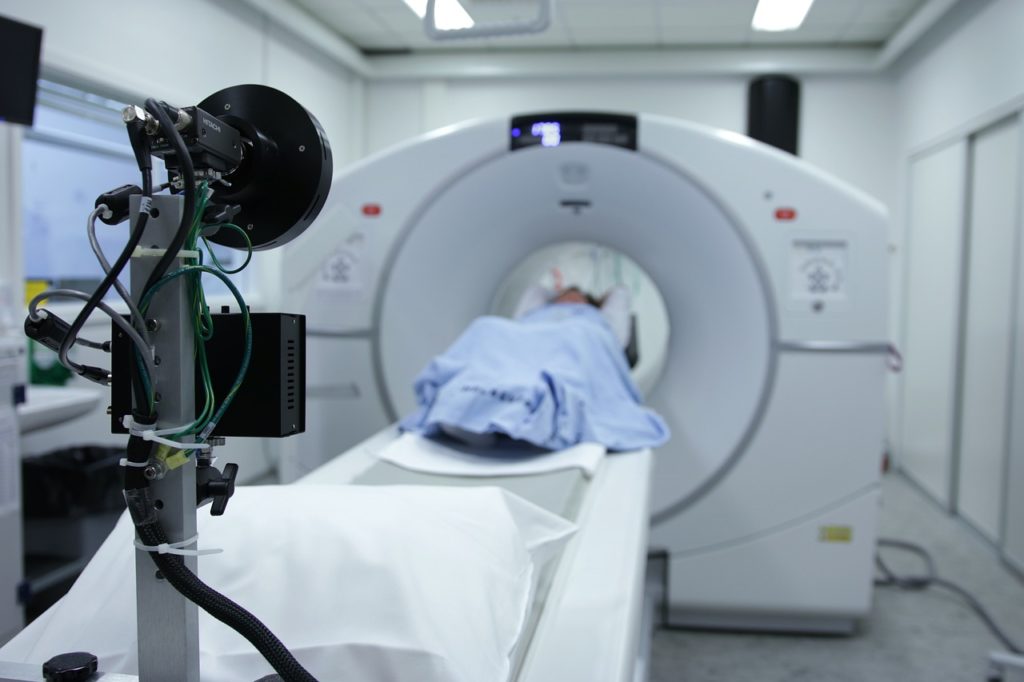
A new study from Brigham and Women’s Hospital, a founding member of the Mass General Brigham healthcare system, suggests that positron emission tomography (PET) brain scans could reveal hidden inflammation in patients with multiple sclerosis (MS) who are being treated with highly effective treatments. The findings were published in Clinical Nuclear Medicine.

“One of the perplexing challenges for clinicians treating patients with MS is after a certain amount of time, patients continue to get worse while their MRIs don’t change,” said lead author Tarun Singhal, MD, MBBS, an associate professor of Neurology in the Brigham’s Department of Neurology and director of the PET Imaging Program in the Ann Romney Center for Neurologic Diseases.  “This is a new approach that is potentially going to be very helpful for the field, for research, and hopefully for clinical use.”

Singhal collaborated with others in the Brigham Multiple Sclerosis Center and the Ann Romney Center. The study started when Singhal noticed that patients treated with the most effective MS treatments were experiencing worsening symptoms. The team has worked for the past eight years on developing an approach of imaging cells called microglia. Microglia are immune cells in the brain that are thought to have a role in MS disease progression but cannot be seen by a routine MRI. The team developed a technique called F18 PBR 06 PET imaging. It involves the injection of a tracer, or dye, that binds to the microglia cells.

Rohit Bakshi, MD, of the Department of Neurology and a co-author on the paper, said increased microglial activity means more atrophy of grey matter in the brain.

“This can affect cognition, movement, fine motor skills, and other aspects of their life,” Bakshi said.

In their paper, the authors describe the term “smouldering” inflammation. Just as a smouldering fire can burn slowly without smoke or flame, smouldering inflammation may linger in patients with MS, driving disease progression and symptoms, even when it cannot be assessed on MRI.

The newly published study involved performing PET scans on 22 people with MS and eight healthy controls. Researchers measured the glial activity load on the PET scans, a new measure developed in Singhal’s lab where lab members looked at the level of smouldering inflammation from microglia in MS patients. They compared those scans to patients’ disability and fatigue levels and not only found that PET scans could show hidden inflammation caused by microglia, but the damage to patients’ brains correlated with the disability and fatigue levels they were experiencing. The researchers were also able to better classify patients with MS between high-efficacy and low-efficacy treatments. Those with low-efficacy treatments had more abnormalities on their PET scans, suggesting more microglial cell activation. Those using high-efficacy treatments had a lower degree of PET abnormality than those on no or low-efficacy treatments but still had an abnormal increase of microglial activation compared to healthy people, suggesting that while high-efficacy treatments helped to reduce neuroinflammation, there was residual inflammation despite treatment, which could account for future worsening and progression independent of relapse activity (PIRA) in these MS patients.

“Our therapies are excellent in that we’ve improved MS patients’ lives,” Bakshi said. “There’s no doubt about that, but we’re still not at the finish line.”

One limitation to the study is the initial group was small. The authors note that PET scans can also be expensive and expose patients to some level of radiation, whereas MRIs do not. Singhal said that radiation could be reduced because of the long half-life and the requirement for a lower administered dose of the F18 PBR06 tracer. The tracer also produces better imaging characteristics than previously used tracers with shorter half-lives.

Bakshi said despite the limitations, the study shines an important light on the power of PET scanning, specifically to find microglial activation.

“This study tells us something new about the disease and may be giving us an important clue as to what is driving disease progression in patients,” he said.

Singhal said before the technique can be used routinely in a clinical setting, it must be validated on a larger sample size. Other longer half-life PET tracers have been approved by the FDA for clinical use, for example, amyloid PET tracers for studying Alzheimer’s disease. If approved, [F-18]PBR06 could also be used to personalize and predict a patient’s treatment course in MS and other brain diseases. However, the authors note that even before approval, [F-18]PBR06 can help advance drug development and perform multicentric clinical trials.

“It’s very exciting that our novel approach worked and correlated so strongly with clinical measures we assessed,” he said. “It means our approach is relevant clinically.”

Chronic pain linked to socioeconomic background – new study

Development of chronic musculoskeletal pain can be influenced by socioeconomics, fear of movement, smoking and poorer support networks, new research shows. 

In a systematic review of current evidence, researchers found that people from a lower socioeconomic background were twice as likely to develop chronic pain following injury.  

Those with a combination of characteristics, including smoking, high level of pain at the time of injury, fear of movement, poorer support networks and a lower level of education or household income, maybe seven times more likely to develop chronic pain after an injury. The results are published in PLOS One. 

Pain is described as ‘acute’ when it has been present for a short period of time – anything that lasts for less than three months after initial injury. Pain is described as chronic when it has been present for longer than three months after initial injury. Chronic musculoskeletal pain affects about 43 per cent of the UK population and is the greatest cause of disability worldwide, often persisting for many years or indefinitely. People with chronic pain often experience poorer quality of life and are also more likely to develop diseases including cancer, cardiovascular diseases and diabetes.  

Current approaches to managing chronic pain focus on physical rehabilitation at the site of the pain, or injury. However, the body’s healing process usually takes place over no longer than three months, suggesting that the reasons for longer-term pain are more complex. 

Lead author Michael Dunn, of the University of Birmingham and St. George’s University Hospitals NHS Foundation Trust, said: “The purpose of acute pain is to alter behaviour to protect the body from harm, but chronic pain persists because of a sensitised nervous system that continues our experience of pain, even after the healing process has completed.”  

This process, the researchers found, is influenced by a range of psychological and social factors and so treatment which focuses solely on the injured body part is often ineffective. 

Mr Dunn continued: “The characteristics that we have identified are related particularly to an individual’s experiences, rather than a type of injury. For that reason, approaches to treating people with musculoskeletal injuries should be more person-centred, focusing on broader biological, psychosocial and social well-being. Put simply, current healthcare approaches do not address all the reasons people do not get better.” 

The researchers also identified other factors related to developing chronic pain, such as lower job satisfaction, stress and depression. These characteristics were supported by lower quality evidence, but are also linked to lower socioeconomic backgrounds. 

“People from lower socioeconomic backgrounds are twice as likely to develop chronic pain after injury. This indicates that not only are current healthcare approaches inadequate, they may also be discriminatory, with current healthcare approaches that are orientated around the injured body part being geared towards those from higher socioeconomic backgrounds who are less likely to experience these psychological or social factors,” said Mr Dunn.  

New research unveils three distinct language comprehension phenotypes in autistic children.

Largest study of language comprehension acquisition in autistic children reveals a more complex development process and advocates for new therapy approaches

According to The National Institute on Deafness and Other Communication Disorders (NIDCD), Autism can affect children’s language development and communication in a variety of ways. While some children with autism may encounter difficulties communicating verbally, others exhibit impressive vocabularies and the ability to speak on specific topic areas in detail. A new study led by Boston University neuroscientist Dr. Andrey Vyshedskiy examines these unique pathways of language development in autistic children. Published in npj Mental Health Research, this research, encompassing data from over 31,000 autistic individuals, is the most extensive on language comprehension acquisition in autism. Dr. Vyshedskiy’s study supports an idea he had more than two decades ago: that language learning for autistic individuals doesn’t happen in a straight line but goes through three different stages. In this Q&A, he discusses the research’s key takeaways and implications for clinicians, educators, and caregivers.

Can you summarize the main insights from this study?

The common intuitive belief is that language comprehension development follows a linear trajectory: children acquire one grammatical rule at a time. Based on a neurological approach, I predicted over 20 years ago that language unfolds in three steps instead of linear development corresponding to three language comprehension mechanisms of increasing complexity. The new study of over 31,000 autistic individuals, published in npj Mental Health Research, validates this prediction. The implications of this discovery are important for philosophy, paleoanthropology, linguistics, clinical medicine, and improving language therapy interventions for children with autism.

What was the traditional understanding of how children develop language, and how does this research challenge that view?

The current practice of characterizing children’s communication ability only regarding their speech (i.e., verbal, nonverbal, or minimally verbal) is insufficient and one-sided. The new results show that communication abilities can vary independent of verbal abilities. For example, nonverbal children with full syntactic language comprehension have a normal ability to communicate, albeit nonverbally, while verbal children lacking syntactic language comprehension do not have a normal ability to communicate by any means. The new study demonstrates that the three identified language comprehension mechanisms are neurologically and clinically distinct from speech ability. A combined two-dimensional language characterization in terms of both language comprehension and verbal level will better identify children’s communication ability and lead to more children reaching their full linguistic potential.

How many people were involved in this study? How did the research team collect and analyze information?

This is the largest study of language comprehension acquisition in autistic children. It included over 31,000 individuals and was conducted by surveying parents via a language therapy app popular among families with children diagnosed with autism and other language deficits.

Based on your research findings, how might clinicians and educators adapt their approaches to assessment and intervention to better address the diverse communication abilities of autistic children?

Language comprehension in children is commonly assessed based on vocabulary. This evaluation method can grossly misrepresent the child’s actual language comprehension progress. Moreover, it encourages therapists to focus on vocabulary training at the expense of exercises that build full language comprehension. The new study provides evidence for creating new assessments evaluating the three mechanisms of language comprehension. These assessments are poised to improve language therapy interventions and enhance outcomes for individuals with language deficits.

What advice would you give to parents or caregivers based on the implications of this research for understanding and fostering language development in autistic children?

Focus all your efforts on improving your child’s language comprehension. Connecting words is more important than building vocabulary. Language comprehension exercises are included in the book I wrote for parents, This Way to Language: Four Things to Do at the First Sign of Autism.

Moving forward, what areas of research or practice do you believe warrant further exploration to better understand and help autistic children with their language development?

Our recent research demonstrates that the critical period for language acquisition is significantly shorter in autistic children compared to typical children. The shortened critical period may be the main culprit preventing autistic children from acquiring full language. These findings suggest that intensive language therapy has to start much earlier in children with suspected language deficits and encourage research into the possibility of pharmacological extension of the critical period for language acquisition.

Groundbreaking study reveals potential diagnostic marker for multiple sclerosis years before symptom onset.

A new study published today in Nature Medicine unveils a significant breakthrough in the understanding and early detection of multiple sclerosis (MS).

Researchers have identified a unique autoantibody signature present in approximately 10% of patients with MS years before the onset of clinical symptoms.

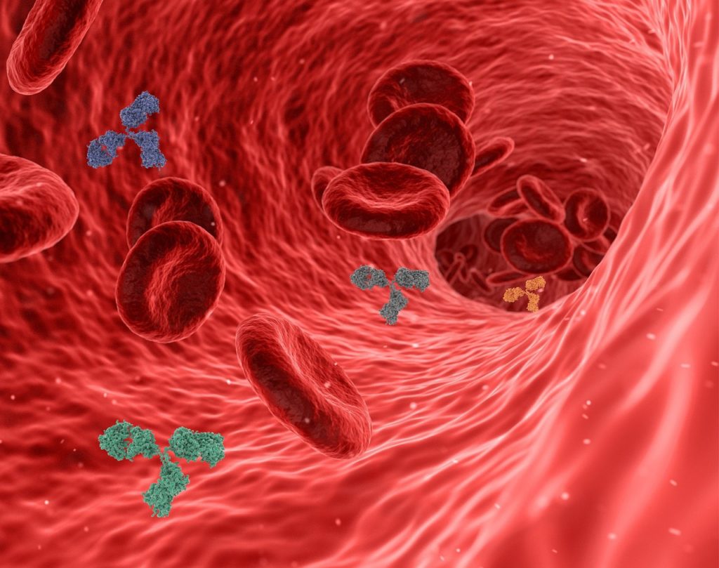
Autoantibodies are antibodies that are supposed to fight off invaders but end up turning against one’s own body, causing problems like autoimmune diseases. Utilizing the U.S. Department of Defense Serum Repository, a cohort encompassing more than 10 million individuals, researchers conducted whole-proteome autoantibody profiling on hundreds of MS patients’ samples collected before and after symptom onset. They discovered a distinct cluster of patients exhibiting an autoantibody signature targeting a common recognizable pattern. Notably, these patients showed antibody reactivity years before developing any MS symptoms and had elevated levels of serum neurofilament light (sNfL), indicating early neuroaxonal injury.

Danillo Augusto, Ph.D., an assistant professor in biology at the University of North Carolina at Charlotte and a co-author of the study, stated, “This study sheds light on the preclinical phase of MS and provides a promising avenue for early detection and intervention. Identifying patients at high risk of developing MS before symptom onset could revolutionize patient care and treatment strategies.”

Further validation of this autoantibody signature was conducted on samples from a separate MS cohort, confirming its high specificity for patients diagnosed with MS. This finding marks a significant milestone in MS research, potentially paving the way for the development of antigen-specific biomarkers for high-risk individuals with clinically or radiologically isolated neuroinflammatory syndromes.