New MRI technique could improve diagnosis and treatment of multiple sclerosis

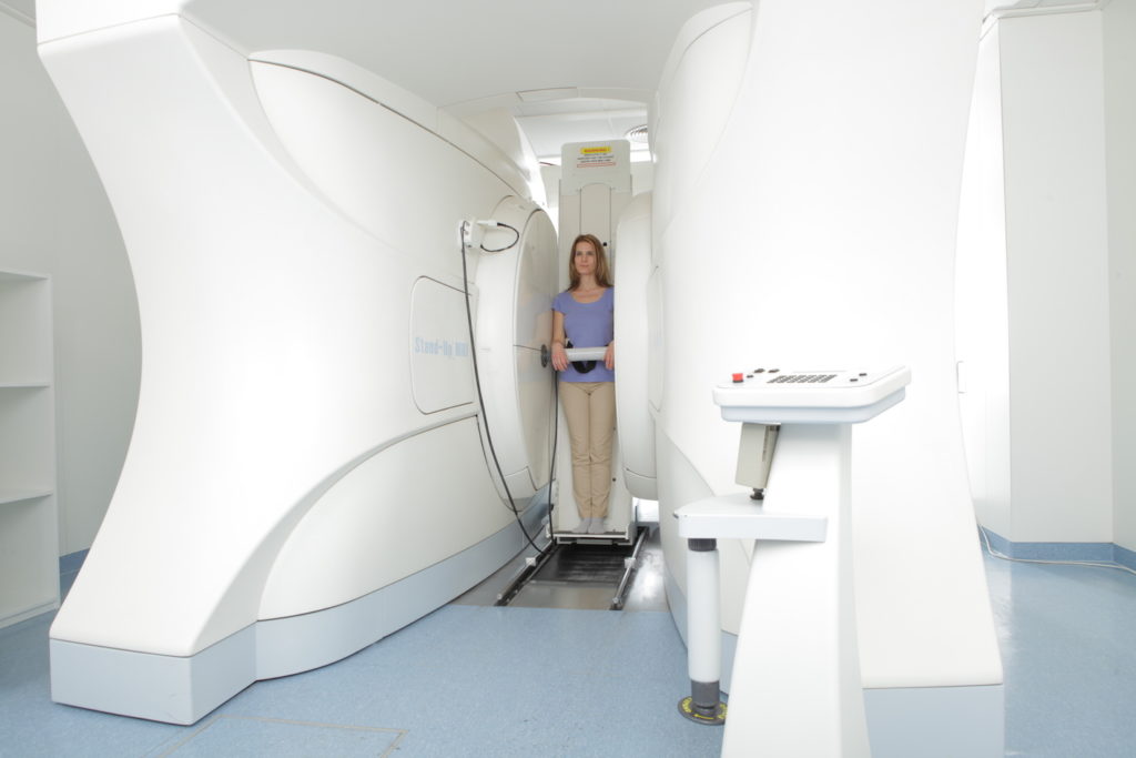
Woman in Medserena Upright Open MRI scanner - no claustrophobia
Woman in Medserena Upright Open MRI scanner – no claustrophobia


It is important that multiple sclerosis (MS) is diagnosed and treated as early as possible in order to delay progression of the disease. The technique of magnetic resonance imaging (MRI) plays a key role in this process. In the search for ever better methods, a new MRI technique has been used at MedUni Vienna as part of a research project that could pave the way to quicker assessment of disease activity in MS. The study was conducted by a research team led by Wolfgang Bogner at MedUni Vienna’s Department of Biomedical Imaging and Image-guided Therapy and was recently published in the leading journal “Radiology“.

Multiple sclerosis is a disease of the central nervous system that manifests itself in changes (lesions) primarily in the brain. As yet, there is no cure for MS, but it can be effectively treated. Early diagnosis is critical to the prognosis, with highly detailed imaging techniques playing a major role. Although conventional MRI can detect brain lesions, scientists are researching methods to detect the changes at an earlier microscopic or biochemical stage. The method known as proton MR spectroscopy has been identified as a promising tool for this purpose.

Using this technique, the research group led by Eva Niess (formerly Heckova) and Wolfgang Bogner from MedUni Vienna’s Department of Biomedical Imaging and Image-guided Therapy, working with scientists from MedUni Vienna’s Department of Neurology, went one step further in their recently published study. The team used MR spectroscopy with a 7-tesla magnet to compare the neurochemical changes in the brains of 65 MS patients with those of 20 healthy controls. This particularly powerful imaging tool was co-developed by MedUni Vienna researchers and has been used for scientific studies, e.g., of the brain, at MedUni Vienna’s Center of Excellence for High-Field MR since it was commissioned in 2008.

Identifying and predicting changes
Using 7-tesla MRI, MedUni Vienna researchers have now been able to identify MS-relevant neurochemicals, i.e. chemicals involved in the function of the nervous system. “This allowed us to visualize brain changes in regions that appear normal on conventional MRI scans,” says study leader Wolfgang Bogner, pointing to one of the study’s main findings. According to the study’s lead author, Eva Niess, these findings could play a significant role in the care of MS patients in the future: “Some neurochemical changes that we’ve been able to visualize with the new technique occur early in the course of the disease and might not only correlate with disability but also predict further disease progression.”

Clinical studies and further developments follow
More research is needed before these findings can be incorporated into clinical applications, explain Niess and Bogner. They say that the results already show 7-tesla spectroscopic MR imaging to be a valuable new tool in the diagnosis of multiple sclerosis and in the treatment of MS patients.

“If the results are confirmed in further studies, this new neuroimaging technique could become a standard imaging tool for initial diagnosis and for monitoring disease activity and treatment in MS patients,” says Wolfgang Bogner, looking to the future. The method is currently only available on the only 7-Tesla MRI scanner in Austria at MedUni Vienna and only for research purposes. However, the scientific team led by Eva Niess and Wolfgang Bogner is working on refining the new method for use in routine clinical MRI scanners.

New MRI technique detects Multiple Sclerosis brain changes earlier

New MRI Technique Detects MS Brain Changes Earlier

(A, B) Sample spectra and abnormal metabolic images of myo-inositol (mI), N-acetylaspartate (NAA), and the ratio of mI to NAA (mI/NAA) together with MRI scans in two participants with multiple sclerosis. Red arrow in A indicates region in normal-appearing white matter with higher signal intensity of mI only. Yellow arrow in B indicates region with higher signal intensity of mI and reduced signal intensity of NAA, where no changes or only diffuse changes are visible at T1-weighted MRI (T1w)/ fluid-attenuated inversion-recovery (FLAIR) imaging, and green arrow indicates white matter lesions, where elevated mI signal intensity extends FLAIR-visible pathologic findings. i.u. = institutional unit, ppm = parts per million, tCho = total choline, tCr = total creatine. CREDIT Radiological Society of North America

A new neuroimaging technique can detect biochemical changes in the brains of people with multiple sclerosis (MS) early in the course of the disease, paving the way for faster treatment evaluation and other potential benefits, according to a study published in the journal Radiology.

MS is a disease of the central nervous system that can cause fatigue, pain and impaired coordination. It affects nearly 3 million people worldwide, and incidence is rising. There is no cure, but physical therapy and medications can slow its progression.

Lesions to the brain’s signal-carrying white matter are the most readily detectable manifestation of MS on MRI. The lesions, linked to the loss of the protective coating around white matter fibers called myelin, represent only macroscopic tissue damage. A means to find changes in the brain at an earlier microscopic or biochemical stage would be beneficial.

An advanced imaging technique known as proton MR spectroscopy is a promising tool in this effort. MR spectroscopy of the brain can detect several metabolites, or substances produced during metabolism, that have potential relevance for MS.

Researchers in Austria used the technique to compare biochemical changes in the brains of 65 people with MS with those of 20 healthy controls. They deployed an MRI scanner with a powerful 7-Tesla (T) magnet.

The results showed reduced levels of an amino acid derivative called N-acetylaspartate (NAA) in patients with MS. Lower levels of NAA have been linked to impaired integrity of neurons in the brain. People with MS also showed elevated levels of myo-inositol (MI), a compound involved in cell signaling. Higher levels are indicative of substantial inflammatory disease activity.

The metabolic alterations in normal-appearing white matter and cortical gray matter were associated with disability.

Researchers said the results show a potential role for 7T MR spectroscopic imaging in visualizing MS pathology beyond demyelinating lesions.

“MRI of neurochemicals enables the detection of changes in the brain of multiple sclerosis patients in regions that appear inconspicuous on conventional MRI,” said study senior author Wolfgang Bogner, Ph.D., from the High Field MR Centre at the Medical University of Vienna in Vienna, Austria. “The visualized changes in neurochemistry of normal-appearing brain tissue correlated with the patients’ disabilities.”

The changes detected by the new imaging technique have significant clinical applications, according to study lead author Eva Heckova, Ph.D., from the High Field MR Centre at the Medical University of Vienna.

“Some neurochemical changes, particularly those associated with neuroinflammation, occur early in the course of the disease and may not only be correlated with disability, but also be predictive of further progression such as the formation of multiple sclerosis lesions,” she said.

While more work is needed to confirm the results, the results support 7T MR spectroscopic imaging as a valuable new aid in the care of people with MS.

“If confirmed in longitudinal clinical studies, this new neuroimaging technique could become a standard imaging tool for initial diagnosis, for disease progression and therapy monitoring of multiple sclerosis patients and, in concert with established MRI, might contribute to neurologists’ treatment strategies,” Dr. Bogner said.

The researchers are working to further improve the image quality of the new technique and fully integrate it for use in routine clinical MRI scanners.

New MRI Technique Detects MS Brain Changes Earlier

Metabolic maps showing the ratio of myo-inositol to N-acetylaspartate (mI/NAA) clearly depict small subcortical or juxtacortical lesions (circles) that appear inconspicuous at T1-weighted MRI (T1w)/fluid-attenuated inversion-recovery (FLAIR) imaging (indicated with arrows in three participants with multiple sclerosis) CREDIT Radiological Society of North America

“In parallel, we will continue our ongoing longitudinal clinical study to validate its ability to detect clinically important pathologic changes in the brain of multiple sclerosis patients and to evaluate the efficacy of different treatment regimens earlier than other currently available clinical tools,” Dr. Heckova said.

High-strength MRI tracks MS progression


Axial 7.0-T T2*-weighted images show examples of leukocortical lesions (white arrows) and intracortical lesions (black arrows) along with juxtacortical and periventricular white matter lesions in different patients with multiple sclerosis (MS). (a) Images of two different brain locations in a 59-year-old man with secondary progressive MS (SPMS). (b) Images of two different brain locations in a 40-year-old woman with SPMS. Radiological Society of North America

The development of scars, or lesions, in the brain’s cortical gray matter is a powerful predictor of neurological disability for people with multiple sclerosis (MS), according to study appearing in the journal Radiology. Researchers said that the findings suggest a role for ultra-high-field-strength MRI in evaluating the progression of MS.

MS is a disease in which the body’s immune system attacks the protective covering surrounding the nerves of the central nervous system. It was once considered a disease of the brain’s white matter, but recent research has shown that cortical lesions, or lesions in the gray matter of the outer layer of the brain, develop even earlier in the course of the disease. These lesions are not easy to see with conventional-strength MRI.

For the new study, researchers tracked MS patients using a 7-Tesla (T) MRI scanner–an extremely powerful machine that has more than twice the magnetic field strength of the more commonly available 3T scanners.

“Because 7T MRI is more sensitive to cortical lesions than lower-field MRI, we can detect many of these lesions that we couldn’t see before and determine if they are strongly correlated with neurological disability and disease progression,” said study senior author Caterina Mainero, M.D., Ph.D., from the Athinoula A. Martinos Center for Biomedical Imaging at Massachusetts General Hospital in Boston. “In this study, we wanted to track the evolution of these lesions and better understand where in the cortex these lesions develop more frequently.”

Dr. Mainero and colleagues followed 20 relapsing-remitting and 13 secondary-progressive MS patients over time, along with 10 age-matched healthy controls. Relapsing-remitting is a type of MS in which the symptoms sometimes improve and sometimes worsen, while secondary-progressive is characterized by more significant disability.

Twenty-five of the MS patients, or 80 percent, developed new cortical lesions, and the 7T MRI detected them more frequently compared to previous studies at lower-field MRI strength. On average, the number of lesions that developed in the cortical region was more than twice the number that developed in the brain’s white matter. The total volume of cortical lesions was a predictor of neurological disability at both baseline and follow-up assessment.

The 7T brain scans showed that the cortical lesions tended to accumulate in grooves on the brain’s surface called sulci.

“We showed that the cortical sulci are the regions where most of these lesions develop,” Dr. Mainero said. “We also found that these lesions can predict disability progression more than white matter lesions, which are the typical lesions of MS we’ve been studying for years.”

While the reasons for the accumulation of lesions in the sulci are not definitively known, researchers note that the flow of cerebrospinal fluid–the fluid that surrounds the brain and spine–is likely to be restricted there. The restricted flow might make the sulci more vulnerable to inflammatory responses.

The results suggest that assessment of cortical lesions should represent a main component in the evaluation of progression of disease burden in MS, Dr. Mainero said.

“This can have a very powerful impact on how we monitor patients with MS,” she said. “We can also use this tool to see how potential treatments can affect the development and evolution of cortical lesions.”

The researchers are looking to replicate their data in larger populations. They also hope to learn more about which patients tend to accumulate more lesions and what factors may be behind the inflammatory response.

“This approach adds another piece to the puzzle of understanding MS,” Dr. Mainero said.

‘A way cool way to be’: Study offers new insights into children with autism


BYU researchers help prepare a child for an MRI scan Bradley Slade

It’s a challenging proposition for anyone: Lie flat on your back, helmet covering half of your face, in a tight-quartered MRI capsule that would make even the non-claustrophobe jittery. Then try not to move for 45 minutes. For children and adolescents with autism and low verbal and cognitive performance (LVCP), that proposition has been nearly impossible without sedation — until now.

Using a range of behavioral support procedures to minimize fear and anxiety, a team of interdisciplinary researchers from Brigham Young University and the University of Utah managed to complete structural and functional MRI scans of 37 children and adolescents with autism, including 17 with less-developed language skills and an average IQ of 54.

“This is such an overlooked group of kids and adults,” said Ryan Kellems, BYU assistant professor of counseling psychology and special education. Kellems also co-authored the study, just published in top academic journal Molecular Autism. “We don’t know very much about their brains because people say they are challenging to study.”

Other researchers have done structural MRIs of individuals with LVCP populations under sedation, but this group, spearheaded by autism pro and BYU assistant professor of school psychology Terisa Gabrielsen, wanted to find a way to watch their brains while they were awake and working. So they created a video explaining the step-by-step fMRI process children could watch at home multiple times, and they provided families with audio files that would help children prepare for the sounds they’d hear in the machine.

When children arrived for their scans, the researchers let them push buttons on the machine so they could see it move up and down. And once the scans began, added Gabrielsen, “if they were getting antsy, we reassured them. We could hold their hand. We could remind them to stop moving. We were focused on giving them more than what they might need to be successful in the scanner.”

With the scans they were able to complete, the team analyzed how those from the LVCP group compared to higher performance children with autism, as well as neurotypical children.

The team found that within the LVCP group, a number of the brain’s networks “just weren’t working in sync as much,” Gabrielsen. “And we also found decreased activity between the left and right hemispheres.”

Another finding practitioners have observed clinically but haven’t yet been able to observe in the brain: network connectivity was higher for the LVCP group than for the neurotypical group.

Explained study co-author Mikle South, BYU associate professor of psychology and neuroscience: “You might see a kid without very much language, and you might guess that his brain is underactive. But sometimes it’s more active. Sometimes the attention is too much, and sometimes the attention is too little. We see different patterns of what the brain is attending to.”

Understanding what’s happening in the brain, how information is being processed and what is or isn’t being attended to, South explained, will allow researchers and practitioners to ultimately help make life better for individuals with autism and their families.

“We don’t want to change the brain, we want to accept folks with autism,” said South, who is also an autism scholar. “This is a way cool way to be, and we just want to help you get along in your world.”

Moving forward, the team would like to do follow up scans with the same children, and also get larger samples of each of the groups: “Getting younger kids, kids with even less language, all of that would be helpful and solidify the findings and tell us more,” said Gabrielsen. “Thanks to the participants in this study, we know how to do it now.”


Does widespread pain stem from the brain? MRI study investigates


New, more powerful MRI scanners may cause fillings to leak mercury

New, more powerful MRI scanners may cause fillings to leak mercury

Pain is the most common reason people seek medical care, according to the National Institutes of Health.

“Sometimes we can easily pinpoint what is causing a person pain,” says Richard Harris, Ph.D., associate professor of anesthesiology and rheumatology at Michigan Medicine. “But, there are still 1 in 5 Americans who suffer from persistent pain that is not easily identifiable.”


Whenever someone experiences pain, they often think about how intense the pain is — but rarely do they also consider how widespread the pain is.

Harris is the senior author on a new study, published in Pain, that sought to find what underlies widespread pain.

“We examined data from the brains of participants in the Multidisciplinary Approach to the Study of Chronic Pelvic Pain Research Network study,” Harris says. “We compared participants with a clinical diagnosis of urological chronic pelvic pain syndrome to pain-free controls and to fibromyalgia patients.”

Harris and colleagues examined if widespread pain, thought to be a marker of centralization in the nervous system, actually originates in the brain.

Functional and structural neuroimaging

The research team used data from 1,079 participants from the MAPP study that included questionnaires capturing their pain severity and function. They were also asked to draw on a body map where they were experiencing pain.

Researchers then had a subset of the participants undergo functional and structural MRIs.


“Surprisingly, many of the individuals, in addition to having pain located in the pelvic region, had pain also widely distributed throughout their body,” Harris says. “Interestingly, when we put these individuals into the brain imaging scanner, we found that those who had widespread pain had increased gray matter and brain connectivity within sensory and motor cortical areas, when compared to pain-free controls.”

Urological chronic pelvic pain syndrome patients with widespread pain showed increased brain gray matter volume and functional connectivity involving the sensorimotor and insular cortices.

“What was surprising was these individuals with widespread pain, although they had the diagnosis of urological chronic pelvic pain, were actually identical to another chronic pain disorder: fibromyalgia,” Harris says.

The team notes the changes in brain gray matter volume and functional connectivity were identical to outcomes present in fibromyalgia patients, but were not seen in the pain-free control group.

“This study represents the fact that pelvic pain patients, a subset of them, have characteristics of fibromyalgia,” Harris says. “Not only do they have widespread pain, but also they have brain markers indistinguishable from fibromyalgia patients.”

Harris and colleagues hope this study provides physicians with the opportunity to look at new ways of treating chronic pain patients — as there might be similarities across pain conditions if both show widespread pain.

“We think that this type of study will help treat these patients because if they have a central nerve biological component to their disorder, they’re much more likely to benefit from targets that affect the central nervous system rather than from treatments that are aimed at the pelvic region,” Harris says.