Chiari malformation – What are the signs and symptoms of Chiari malformations?

Chiari malformation - - what are the signs and symptoms?

Chiari malformation – – what are the signs and symptoms?

A Chiari malformation, previously called an Arnold-Chiari malformation, is where the lower part of the brain pushes down into the spinal canal.

There are four main types, but type 1, called Chiari I, is the most common.

In someone with Chiari I, the lowest part of the back of the brain extends into the spinal canal. This can put pressure on the brainstem, spinal cord, and obstruct the flow of fluid.

This page focuses on Chiari I malformations.

Are Chiari I malformations serious?

The severity of Chiari malformations can vary from person to person, but generally:

Chiari I malformations aren’t considered life-threatening

some people experience painful headaches, movement problems and other unpleasant symptoms (see below), but many people won’t have any symptoms

there’s a chance of developing syringomyelia (where a fluid-filled cavity called a syrinx develops in the spinal cord), which can damage the spinal cord if not treated promptly

surgery can usually stop the symptoms getting worse and can sometimes improve them, although some problems may remain

Talk to your doctor about what the condition means, what the implications may be for your health and what treatment you may need.

Symptoms of Chiari I malformations

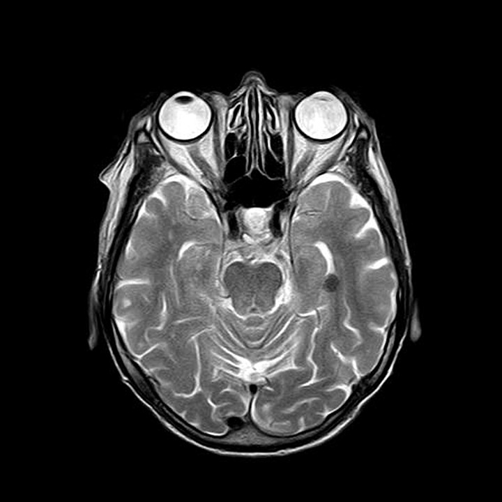
Many people with a Chiari I malformation will not have any symptoms. Sometimes they’re only found after an (MRI) scan of the brain is carried out for another reason.

If symptoms do develop, they can include:

headaches – these are usually felt at the back of the head and may be brought on or made worse by coughing, straining, sneezing or bending over

neck pain

dizziness and balance problems

muscle weakness

numbness or tingling in the arms or legs

blurred vision, double vision and sensitivity to light

swallowing problems

hearing loss and tinnitus

feeling and being sick

difficulty sleeping (insomnia) and depression

If you develop syringomyelia, you may also experience problems using your hands, difficulty walking, pain, and problems with bladder or bowel control.

If you’ve been diagnosed with a Chiari malformation, you should contact your doctor for advice if you develop any new symptoms or your symptoms worsen.

Treatments for Chiari I malformations

Treatment for Chiari I malformation depends on whether you have any symptoms and how severe they are. You might not need any treatment if you don’t have any symptoms.

Painkillers can help relieve any headaches and neck pain.

If your headaches are severe or you have problems caused by the pressure on your spinal cord (such as movement difficulties), surgery may be recommended.

Surgery

The main operation for Chiari malformation is called decompression surgery.

Under general anaesthetic a cut is made at the back of your head and the surgeon removes a small piece of bone from the base of your skull. They may also remove a small piece of bone from the top of your spine.

This will help reduce the pressure on your brain and allow the fluid in and around your brain and spinal cord to flow normally. Read an NHS leaflet about decompression for Chiari malformation (PDF, 111kb).

Other procedures that may be necessary include:

Endoscopic third ventriculostomy (ETV) – a small hole is made in the wall of one of the cavities of the brain, releasing trapped fluid. See treating hydrocephalus for more information.

Ventriculoperitoneal shunting – a small hole is drilled into the skull and a thin tube called a catheter is passed into the brain cavity to drain trapped fluid and relieve the pressure. See treating hydrocephalus for more information.

Untethering – some children with a type 1 Chiari malformation have a tethered spinal cord, which means it is abnormally attached within the spine. Untethering involves separating the spinal cord and releasing tension in the spine. Read an NHS leaflet on tethered spinal cord (PDF, 193kb).

Spinal fixation – some people with Chiari I will have a hypermobility syndrome, such as Ehlers-Danlos syndrome, and may require surgery to stabilise their spine.

The aim of surgery is to stop existing symptoms getting any worse. Some people also experience an improvement in their symptoms, particularly their headaches.

However, surgery sometimes results in no improvement or symptoms getting worse. There’s also a small risk of serious complications, such as paralysis or a stroke.

Talk to your surgeon about the different surgical options and what the benefits and risks of each are.

Causes of Chiari I malformations

The exact cause of Chiari I malformations is unknown. It tends to be present from birth, but is normally only found in adulthood when symptoms develop or when an MRI scan is done.

Many cases are thought to be the result of part of the skull not being large enough for the brain.

Chiari I malformations can also develop in people with a tethered spinal cord, a build-up of fluid on the brain (hydrocephalus), and some types of brain tumour.

Chiari malformations can sometimes run in families. It’s possible that some children born with it may have inherited a faulty gene that caused problems with their skull development.

But the risk of passing a Chiari malformation on to your child is very small. And remember: even if your children do inherit it, they may not experience symptoms.

New, more powerful MRI scanners may cause fillings to leak mercury

New, more powerful MRI scanners may cause fillings to leak mercury

New, more powerful MRI scanners may cause fillings to leak mercury

“Metal fillings ‘leak mercury after scan’,” reports BBC News. Experiments found teeth with fillings put through new higher powered MRI scanners released more mercury into a test tube of artificial saliva than teeth exposed to conventional lower powered scanners.

Silver-coloured amalgam, which contains mercury, is widely used for fillings in the UK. Although amalgam fillings can release low levels of mercury vapour, particularly when they’re put in or removed, there’s no evidence that exposure to mercury from amalgam fillings has any harmful effects on health.

MRI scans may heat up the dental amalgam, which might increase leakage of mercury vapour. Previous studies using standard power MRI scanners haven’t found any evidence of an increase in mercury vapour.

But the researchers looked at what are known as 7.0-Tesla MRI scanners. These produce much more powerful magnetic waves than conventional MRI scanners, so they’re currently mainly used for research and during clinical trials. At the time of writing, they’re rarely used for standard diagnostic procedures.

This experimental study used test tubes with extracted teeth and artificial saliva. We don’t know if the results would be the same when people with fillings have MRI scans, or whether mercury vapour released in this way is absorbed by the body in a harmful way.

The study is useful, as it’s likely the cost of 7.0-Tesla MRI scanners will drop in the years to come and they may become more widely used.

Where did the story come from?

The study was carried out by researchers from Akdeniz University and Kirikkale University in Turkey.

It was funded by Kirikkale University Scientific Research Projects Coordination Unit.

The study was published in the peer-reviewed journal Radiology on an open access basis, so it’s free to read online.

The Mail Online played up the potential dangers of mercury, stating: “High levels of mercury in the blood can cause brain damage such as paralysis or slow reflexes, may increase the risk of heart attack or heart disease, and can reduce men’s sperm counts or increase the risk of a woman having a deformed baby.”

While this is correct, the study doesn’t tell us whether the mercury released from the fillings could make its way into the blood at a high enough level to cause these problems.

BBC News and The Independent carried more balanced stories.

What kind of research was this?

This experimental laboratory research, carried out in test tubes (ex-vivo) rather than in the human body. It is early-stage research to look for potential problems which might need further investigation.

The next-generation of 7 Telsa MRI scanners have the advantage of providing a much more detailed image. As BBC News put it in a 2016 report: “going from an ordinary TV picture to HD”.

A potential concern is that these new types of scanner generate a much greater magnetic field than the kind of MRI scanner you would find in your local hospital.

What did the research involve?

Researchers used 60 teeth which had been extracted for clinical reasons. All were given standard dental amalgam fillings. The teeth were then randomly divided into 3 groups of 20. Nine days after the fillings were done, teeth in each group were put into test tubes with artificial saliva.

1 group was stored for 24 hours

1 group was exposed to standard 1.5 T (Tesla) MRI for 20 minutes, then stored for 24 hours

1 group was exposed to high powered 7T MRI for 20 minutes, then stored for 24 hours

After 24 hours, all the teeth were taken out of the tubes and the artificial saliva was analysed for mercury concentration. The researchers compared the concentration between the 3 groups.

What were the basic results?

Mean mercury levels in the artificial saliva were:

141 micrograms per litre (plus or minus 152) for teeth not exposed to MRI

172 micrograms per litre (plus or minus 60) for teeth exposed to standard MRI

673 micrograms per litre (plus or minus 179) for teeth exposed to high powered MRI

The differences between the control group and the group exposed to standard MRI were so small they could have come about by chance.

How did the researchers interpret the results?

The researchers said: “We concluded that, 9 days after dental restoration, exposure to 7.0T MRI was associated with mercury release from dental amalgam in an ex-vivo [out of the body, ie test tube] setting.”

Conclusion

Sensationalist headlines such as the Mail Online’s: “High-strength MRI scanners may release toxic mercury from dental fillings putting people at risk of brain damage, heart attack or infertility” can cause panic, but in this case, there is little to worry about for most people. This is an experimental study, carried out in test tubes not in people, using a technology that only a handful of hospitals in the UK have access to. 7T MRI has only recently been approved for use, and only a few research hospitals have the new scanners.

There have been panics about mercury in fillings for many years now, with no evidence to show that they actually cause harm. The low levels of vapour release usually happen when they are first put in (until the amalgam has hardened and sealed the mercury inside) or if they are taken out. Taking out mercury fillings to replace with alternative fillings (except when the fillings are worn out or broken and need replacing) is likely to do more harm than good.

The standard MRI scans that most people have do not seem to increase the release of mercury vapour. The study suggests this may be more of a problem with the new high-powered scans, and this should be further investigated.

However, the study has limitations:

it cannot tell us whether the mercury found in artificial saliva would be absorbed into human tissue, as the study was carried out in test tubes not in people

the fillings were not polished after being made, as is usually done in practice, which might have caused an increase in release of mercury

the saliva samples were tested just once so we don’t know whether levels of mercury would have reduced over time

we don’t know the effect of repeated MRI scans

In the unlikely event that you need a high-powered MRI scan (for example you are invited to take part in a clinical trial) and have amalgam fillings, it may be worth discussing the pros and cons with doctors and your dentist. They should be able to provide advice about whether the benefits of the high-power scan outweigh any possible risks.

Analysis by Bazian
Edited by NHS Choices

How an MRI scan is performed

MRI scan

MRI scan

A magnetic resonance imaging (MRI) scan is a painless procedure that lasts 15 to 90 minutes, depending on the size of the area being scanned and the number of images being taken.

Before the scan

On the day of your MRI scan, you should be able to eat, drink and take any medication as usual, unless advised otherwise.

In some cases, you may be asked not to eat or drink anything for up to four hours before the scan, and sometimes you may be asked to drink a fairly large amount of water beforehand. This depends on the area being scanned.

When you arrive at the hospital, you’ll usually be asked to fill in a questionnaire about your health and medical history. This helps the medical staff to ensure you have the scan safely. Read more about who can and can’t have an MRI scan.

Once you’ve completed the questionnaire, you’ll usually be asked to give your signed consent for the scan to go ahead.

As the MRI scanner produces strong magnetic fields, it’s important to remove any metal objects from your body, including:

watches

jewellery, such as earrings and necklaces

piercings, such as ear, nipple and nose rings

dentures (false teeth)

hearing aids

wigs (some wigs contain traces of metal)

Any valuables can usually be stored in a secure locker.

Depending on which part of your body is being scanned, you may need to wear a hospital gown during the procedure. If you do not need to wear a gown, you should wear clothes without metal zips, fasteners, buttons, underwire (bras), belts or buckles.

Contrast dye

Some MRI scans involve having an injection of contrast dye. This makes certain tissues and blood vessels show up more clearly and in greater detail.

It’s possible for contrast dye to cause tissue and organ damage in people with severe kidney disease. If you have a history of kidney disease, you may be given a blood test to determine how well your kidneys are functioning and whether it’s safe to proceed with the scan.

You should also let the staff know if you have a history of allergic reactions or any blood clotting problems before having the injection.

Anaesthesia and sedatives

An MRI scan is a painless procedure, so anaesthesia (painkilling medication) isn’t usually required. If you’re claustrophobic, you can ask for a mild sedative to help you relax – you should ask your GP or consultant well in advance of having the scan.

If you decide to have a sedative during the scan, you’ll need to arrange for a friend or family member to drive you home afterwards, as you won’t be able to drive for 24 hours.

General anaesthetic is often used when young children and babies have an MRI scan. This is because it’s very important to stay still during the scan, which young children and babies are often unable to do when they’re awake.

During the scan

An MRI scanner is a short cylinder that’s open at both ends. You’ll lie on a motorised bed that’s moved inside the scanner. You’ll enter the scanner either head first or feet first, depending on the part of your body being scanned.

In some cases, a frame may be placed over the body part being scanned, such as the head or chest. This frame contains receivers that pick up the signals sent out by your body during the scan and it can help to create a better-quality image.

A computer is used to operate the MRI scanner, which is located in a different room to keep it away from the magnetic field generated by the scanner.

The radiographer operates the computer, so they’ll also be in a separate room to you. However, you’ll be able to talk to them, usually through an intercom, and they’ll be able to see you at all times on a television monitor.

While you’re having your scan, a friend or family member may be allowed to stay in the room with you. Children can usually have a parent with them. Anyone who stays with you will be asked whether they have a pacemaker or any other metal objects in their body. They’ll also have to follow the same guidelines regarding clothing and removing metallic objects.

To avoid the images being blurred, it’s very important to keep the part of your body being scanned still throughout the whole of the scan until the radiographer tells you to relax.

A single scan may take from a few seconds to three or four minutes. You may be asked to hold your breath during short scans. Depending on the size of the area being scanned and how many images are taken, the whole procedure will take 15 to 90 minutes.

The MRI scanner will make loud tapping noises at certain times during the procedure. This is the electric current in the scanner coils being turned on and off. You’ll be given earplugs or headphones to wear.

You’re usually able to listen to music through headphones during the scan if you want to, and in some cases you can bring your own CD.

You’ll be moved out of the scanner when your scan is over.

After the scan

An MRI scan is usually carried out as an outpatient procedure. This means you won’t need to stay in hospital overnight. After the scan, you can resume normal activities immediately.

However, if you’ve had a sedative, a friend or relative will need to take you home and stay with you for the first 24 hours. It’s not safe to drive, operate heavy machinery or drink alcohol for 24 hours after having a sedative.

Your MRI scan needs to be studied by a radiologist (a doctor trained in interpreting scans and X-rays) and possibly discussed with other specialists. This means it’s unlikely you’ll know the results of your scan immediately.

The radiologist will send a report to the doctor who arranged the scan, who will discuss the results with you. It usually takes a week or two for the results of an MRI scan to come through, unless they’re needed urgently.

World Radiography Day – A Patient’s Guide to Medical Imaging

World Radiography Day is celebrated on 8 November each year. The date marks the anniversary of the discovery of x-rays by Wilhelm Roentgen in 1895.

Radiographers worldwide can use the day and the days around the date to promote radiography as a career, as a vital contribution to modern healthcare and as a chance to increase public awareness of diagnostic imaging and radiation therapy.

Do we thought it would be useful to share this brief guide to medical imaging which goes from x-rays to MRI scans.

A Patients Guide to Medical Imaging

From Visually.

Getting through an MRI scan. Hints and tips from our readers.

What is it like to have an MRI scan?

What is it like to have an MRI scan?

Over the years there has been a fair bit of discussion of MRI scan both on this blog and our various Facebook pages. For example MRI scans are often used as a test for multiple sclerosis.

MRI is an abbreviation for Magnetic Resonance Imaging. In simple terms the UK’s NHS describes the procedure as follows “Magnetic resonance imaging is a type of scan that uses strong magnetic fields and radio waves to produce detailed images of the inside of the body. An MRI scanner is … a large tube that contains powerful magnets”. They continue with the following description ” During an MRI scan, you lie on a flat bed that is moved into the scanner.”

A radiographer controls “the scanner using a computer, which is in a different room to keep it away from the magnetic field generated by the scanner. You will be able to talk to the radiographer through an intercom and they will be able to see you on a television monitor throughout the scan. At certain times during the scan, the scanner will make loud tapping noises. This is the electric current in the scanner coils being turned on and off. You will be given earplugs or headphones to wear. It is very important that you keep as still as possible during your MRI scan. The scan will last between 15 and 90 minutes, depending on the size of the area being scanned and how many images are taken.”

Doesn’t sound much fun does it?

So we thought it would be useful to get our readers to share their tips and hints for a better MRI experience.

” I have 2 a year one head only takes about 30 mins. Head, neck and spine takes about 1.5 hours and can be quite painful. I can hardly walk when I get out and sleep most of the day away when I get home… I normally just take 2 ibuprofen when I leave for the hospital just to take the pain edge off….and I can also ask them to stop if I need adjusting or need to move for a few mins….”

” I always ask for pillow rolls at my neck, my legs. That has always helps.”

“I always have ” something to help me sleep “before the one and a half hour scan so I usually sleep through it…. The pain at the back of my head and the whole experience becomes bearable. I don’t like breaking the scan into smaller pieces cuz it means I have to wait at the hospital all day”

“Claustrophobic also. They give me versed to knock me out. Usually I wake up half way thru and they then give me more.”

“try the T3 MRI.itscmuch better.butvyou should take something to keep you calm”

“MRI’s are not fun usually if you ask your Dr/nurse they are able to give you something IV to clam you down. If you’ve just been diagnosed MRI’s are very important so your Dr can tell were your lesions are then in the future your MRI’s will have to be compared. Like this past January I had a chronic migraine that I just couldn’t get under control at home so I had to go the hospital for a few days so I could get some IV medication for pain control my neurologist ordered an MRI just to make sure the migraine was from my MS. Guess what! Some of my lesion were not as bright as what they on my last MRI. So you cam see how important they are. Sometimes you have to just grin and bear it”

“i would ask for it to be broken up. I had back surgery in 06 on L5S1 and it hurts me worse now…It is very hard for me to lay there for 15-20mins with the wedge then the.vertigo that i get when trying to set up is horrific as well….gl”

“I am unable to lay still for long periods of time without spasms so I am stuck taking a double dose

Radiologist Image courtesy of imagerymajestic at FreeDigitalPhotos.net

Radiologist Image courtesy of imagerymajestic at FreeDigitalPhotos.net

20 minutes before and tend to fall asleep during. I also get an extra roll under my neck. My MRI team (after you have x amount of MRI’s you get to know the techs) allow me to sit up between the different ones. They do my brain – stretch – cervical spine – stretch – thoracic spine – stretch – lumbar and finally done. And also, maybe get the different ones on different days. I also tell them not to talk to me and just let me know if when they are pulling me out. I jumped every time they would tell me what was going on. I could meditate with the music to stay relaxed. Scaring me with their instructions tightened up my muscles. Only when they had to give instructions and I also told them to use a quieter voice with me. Dumb huh?”

“Thinking of pleasant thoughts helps to relax you and deal with the pain. I too suffer with the pain, hang in there”

“Ask them to take a break so you can at least sit up for awhile”

All bits of good advice for those about to have an MRI scan.

Do you have any?

Feel free to share in the comments section below. You might be interested in using some of the following questions to frame your response:-

1) What is your main medical condition?
2) What was the main reason for having an MRI scan?
3) How many times a year do you have a scan and how long does it take?
4) What is it like to have an MRI scan?
5) What tips or advice would you give to somebody about to have their first MRI scan?

Thanks very much in advance!