Jaundice – what are the causes of Jaundice?

Jaundice

Jaundice

Jaundice is caused by a build-up of a substance called bilirubin in the blood and tissues of the body.

Any condition that disrupts the movement of bilirubin from the blood to the liver and out of the body can cause jaundice.

Bilirubin

Bilirubin is a waste product created when red blood cells break down. It’s transported in the bloodstream to the liver, where it’s combined with a digestive fluid called bile.

This eventually passes out of the body in urine or stools. It’s bilirubin that gives urine its light yellow colour and stools their dark brown colour.

Types of jaundice

There are three types of jaundice, depending on what’s affecting the movement of bilirubin out of the body.

Pre-hepatic jaundice

Pre-hepatic jaundice occurs when a condition or infection speeds up the breakdown of red blood cells. This causes bilirubin levels in the blood to increase, triggering jaundice.

Causes of pre-hepatic jaundice include:

malaria – a blood-borne infection spread by mosquitoes

sickle cell anaemia – an inherited blood disorder where the red blood cells develop abnormally; it’s most common among black Caribbean, black African and black British people

thalassaemia – similar to sickle cell; it’s most common in people of Mediterranean, Middle Eastern and, in particular, South Asian descent

Crigler-Najjar syndrome – a genetic syndrome where an enzyme needed to help move bilirubin out of the blood and into the liver is missing

hereditary spherocytosis – a genetic condition that causes red blood cells to have a much shorter life span than normal

Intra-hepatic jaundice

Intra-hepatic jaundice happens when a problem in the liver – for example, damage due to infection or alcohol, disrupts the liver’s ability to process bilirubin.

Causes of intra-hepatic jaundice include:

the viral hepatitis group of infections – hepatitis Ahepatitis B and hepatitis C

alcoholic liver disease – where the liver is damaged as a result of drinking too much alcohol

leptospirosis – a bacterial infection that’s spread by animals, particularly rats

glandular fever – a viral infection caused by the Epstein-Barr virus

drug misuse – leading causes are ecstasy and overdoses of paracetamol

primary biliary cirrhosis – a rare condition that causes progressive liver damage

Gilbert’s syndrome – a common genetic syndrome where the liver has problems breaking down bilirubin at a normal rate

liver cancer – a rare and usually incurable cancer that develops inside the liver

exposure to substances known to be harmful to the liver – such as phenol (used in the manufacture of plastic) or carbon tetrachloride (widely used in the past in processes such as refrigeration, although now its use is strictly controlled)

autoimmune hepatitis – a rare condition where the immune system starts to attack the liver

primary sclerosing cholangitis – a rare type of liver disease that causes long-lasting (chronic) inflammation of the liver

Dubin-Johnson syndrome – a rare genetic syndrome where the liver is unable to move bilirubin out of the liver

Post-hepatic jaundice

Post-hepatic jaundice is triggered when the bile duct system is damaged, inflamed or obstructed, which results in the gallbladder being unable to move bile into the digestive system.

Causes of post-hepatic jaundice include:

gallstones – obstructing the bile duct system

pancreatic cancer

gallbladder cancer or bile duct cancer

pancreatitis – inflammation of the pancreas, which can either be acute pancreatitis (lasting for a few days) or chronic pancreatitis (lasting for many years)

Some causes of jaundice are common, such as hepatitis and gallstones, whereas other causes, such as Crigler-Najjar syndrome and Dubin-Johnson syndrome, are much rarer.

Acute cholecystitis – find out more about the signs and symptoms of swelling of the gallbladder.

Acute cholecystitis

Acute cholecystitis

Acute cholecystitis is swelling (inflammation) of the gallbladder. It is a potentially serious condition that usually needs to be treated in hospital.

The main symptom of acute cholecystitis is a sudden sharp pain in the upper right side of your tummy (abdomen) that spreads towards your right shoulder.

The affected part of the abdomen is usually extremely tender, and breathing deeply can make the pain worse.

Unlike some others types of abdominal pain, the pain associated with acute cholecystitis is usually persistent, and doesn’t go away within a few hours.


Some people may additional symptoms, such as:

  • a high temperature (fever)
  • nausea and vomiting
  • sweating
  • loss of appetite
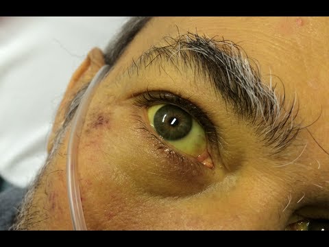
  • yellowing of the skin and the whites of the eyes (jaundice)
  • a bulge in the abdomen

When to seek medical advice

You should see your GP as soon as possible if you develop sudden and severe abdominal pain, particularly if the pain lasts longer than a few hours or is accompanied by other symptoms, such as jaundice and a fever.

If it’s not possible to contact your GP immediately, phone your local out-of-hours service or call NHS 111 for advice.

It’s important for acute cholecystitis to be diagnosed as soon as possible, because there is a risk that serious complications could develop if the condition is not treated promptly (see below).

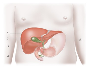
What causes acute cholecystitis?

The causes of acute cholecystitis can be grouped into two main categories: calculous cholecystitis and acalculous cholecystitis.

Calculous cholecystitis

Calculous cholecystitis is the most common, and usually less serious, type of acute cholecystitis. It accounts for around 95% of all cases.

Calculous cholecystitis develops when the main opening to the gallbladder, called the cystic duct, gets blocked by a gallstone or by a substance known as biliary sludge.

Biliary sludge is a mixture of bile (a liquid produced by the liver that helps digest fats) and small crystals of cholesterol and salt.

The blockage in the cystic duct results in a build-up of bile in the gallbladder, increasing the pressure inside it and causing it to become inflamed. In around one in every five cases, the inflamed gallbladder also becomes infected by bacteria.

Acalculous cholecystitis

Acalculous cholecystitis is a less common, but usually more serious, type of acute cholecystitis. It usually develops as a complication of a serious illness, infection or injury that damages the gallbladder.

Acalculous cholecystitis is often associated with problems such as accidental damage to the gallbladder during major surgery, serious injuries or burns, blood poisoning (sepsis), severe malnutrition or AIDS.

Who is affected

Acute cholecystitis is a relatively common complication of gallstones.

It is estimated that around 10-15% of adults in the UK have gallstones. These don’t usually cause any symptoms, but in a small proportion of people they can cause infrequent episodes of pain (known as biliary colic) or acute cholecystitis.

In England, around 28,000 cases of cholecystitis were reported during 2012-13.

Diagnosing cholecystitis

To diagnose acute cholecystitis, your GP will examine your abdomen.

They will probably carry out a simple test called Murphy’s sign. You will be asked to breathe in deeply with your GP’s hand pressed on your tummy, just below your rib cage.

Your gallbladder will move downwards as your breathe in and, if you have cholecystitis, you will experience sudden pain as your gallbladder reaches your doctor’s hand.

If your symptoms suggest you have acute cholecystitis, your GP will refer you to hospital immediately for further tests and treatment.

Tests you may have in hospital include:

  • blood tests to check for signs of inflammation in your body
  • an ultrasound scan of your abdomen to check for gallstones or other signs of a problem with your gallbladder

Other scans – such as an X-ray, a computerised tomography (CT) scan or a magnetic resonance imaging (MRI) scan – may also be carried out to examine your gallbladder in more detail if there is any uncertainty about your diagnosis.

Treating acute cholecystitis

If you are diagnosed with acute cholecystitis, you will probably need to be admitted to hospital for treatment.

Initial treatment

Initial treatment will usually involve:

  • fasting (not eating or drinking) to take the strain off your gallbladder
  • receiving fluids through a drip directly into a vein (intravenously) to prevent dehydration
  • taking medication to relieve your pain

If you have a suspected infection, you will also be given antibiotics. These often need to be continued for up to a week, during which time you may need to stay in hospital or you may be able to go home.

With this initial treatment, any gallstones that may have caused the condition usually fall back into the gallbladder and the inflammation often settles down.

Surgery

In order to prevent acute cholecystitis recurring, and reduce your risk of developing potentially serious complications, the removal of your gallbladder will often be recommended at some point after the initial treatment. This type of surgery is known as a cholecystectomy.

Although uncommon, an alternative procedure called a percutaneous cholecystostomy may be carried out if you are too unwell to have surgery. This is where a needle is inserted through your abdomen to drain away the fluid that has built up in the gallbladder.

If you are fit enough to have surgery, your doctors will need to decide when the best time to remove your gallbladder may be. In some cases, you may need to have surgery immediately or in the next day or two, while in other cases you may be advised to wait for the inflammation to fully resolve over the next few weeks.

Surgery can be carried out in two main ways:

  • laparoscopic cholecystectomy – a type of keyhole surgery where the gallbladder is removed using special surgical instruments inserted through a number of small cuts (incisions) in your abdomen
  • open cholecystectomy – where the gallbladder is removed through a single, larger incision in your abdomen

Although some people who have had their gallbladder removed have reported symptoms of bloating and diarrhoea after eating certain foods, you can lead a perfectly normal life without a gallbladder.

The organ can be useful but it’s not essential, as your liver will still produce bile to digest food.

Read more about recovering from gallbladder removal.

Possible complications

Without appropriate treatment, acute cholecystitis can sometimes lead to potentially life-threatening complications.

The main complications of acute cholecystitis are:

  • the death of the tissue of the gallbladder, called gangrenous cholecystitis, which can cause a serious infection that could spread throughout the body
  • the gallbladder splitting open, known as a perforated gallbladder, which can spread the infection within your abdomen (peritonitis) or lead to a build-up of pus (abscess)

In about one in every five cases of acute cholecystitis, emergency surgery to remove the gallbladder is needed to treat these complications.

Preventing acute cholecystitis

It’s not always possible to prevent acute cholecystitis, but you can reduce your risk of developing the condition by cutting your risk of gallstones.

One of the main steps you can take to help lower your chances of developing gallstones is adopting a healthy, balanced diet and reducing the number of high-cholesterol foods you eat, as cholesterol is thought to contribute to the formation of gallstones.

Being overweight, particularly being obese, also increases your risk of developing gallstones. You should therefore control your weight by eating a healthy diet and exercising regularly.

However, low-calorie, rapid weight loss diets should be avoided, because there is evidence they can disrupt your bile chemistry and actually increase your risk of developing gallstones. A more gradual weight loss plan is best.

Read more about preventing gallstones.

[Original article on NHS Choices website]

Cholangiocarcinoma Awareness Month – Do you know the signs and symptoms of bile duct cancer?


Cholangiocarcinoma Awareness Month - Bile Duct Cancer Awareness

Cholangiocarcinoma Awareness Month – Bile Duct Cancer Awareness

This month as you can see is Cholangiocarcinoma Awareness Month. Or translated from medicalese it is Bile Duct Cancer Awareness Month.

Bile duct cancer is considered a rare form of cancer with only 1,000 people being diagnosed in the UK each year. It has been suggested that generally only 10-20 per million will develop the condition each year.

As our contribution to raising awareness of Cholangiocarcinoma we thought we would share some of the signs and symptoms that a person may have bile duct cancer.

It should be noted that symptoms only show themselves when the cancer is more advance. This is often when the bile duct is blocked.

The main signs and symptoms include

a) Jaundice
b) Itchy skin
c) Weight loss which is unexpected
d) Light colored or greasy poos
e) Belly pain
f) Dark colored urine.

If you have these symptoms it is important you contact your doctor as soon as possible!


Glandular fever! What is glandular fever and what are your experiences with glandular fever? And more on the Epstein-Barr virus

Glandular Fever

Glandular Fever

“Your glands are up!”

How many of us can remember that phrase from our childhoods.  Indeed in each year, according to the UK’s National Health Service 1 in 200 people will contract glandular fever.  That being said, most people who get glandular fever are in their late teens and early twenties.

When I was at school one of my fellow students had a bout of glandular fever resulting in weeks if not months off sick.

So I felt that it could well be useful to produce a brief guide to glandular fever as part of our series of informational blogs.  As with all our blogs your participation is most welcome.  It would be great to hear about your experiences of glandular fever and its impact upon your life and health.  This will, we hope, provide support for others in a similar situation.

The majority of people who develop glandular fever do so in a period of around two months after contracting the Epstein-Barr virus.  This is perhaps the most common virus which has been covered in a previous blog here https://patienttalk.org/calling-everyone-with-an-autoimmune-condition-have-you-ever-been-infected-with-the-epstein-barr-virus/.  I think the comments section is of particular value.

The main symptoms  of glandular fever are:-

1)                       Fever.  As the name suggests of course.  In this case it is likely to be over 38ºC or                           100.4ºF (in old money).

2)                       Swollen nodes or glands in the neck.  Hence the name glandular fever.

3)                       Sore throat.

4)                       Fatigue.  You can read more about the impact of fatigue by checking out our recent patient experience blog.  https://patienttalk.org/fatigue-like-wet-cement-exploring-the-difference-between-tiredness-and-fatigue/

In some cases there are a number of rarer symptoms.  These can include jaundice and swollen adenoids.  Jaundice is more common with people in the older age brackets who contract glandular fever.

Normally the infection lasts about two or three weeks, starting to get better after around one week albeit slowly.  That being said the fatigue may last for up to six months after the other symptoms have disappeared.

In milder forms of the fever treatments are normally painkillers which also help fight the inflammation.  In more serious cases hospitalisation may be required.

It is worth noting that there does seem to be a link between Epstein–Barr viral infection and contracting a number autoimmune conditions and other illnesses.   In particular Parkinson’s disease,  Lupus (https://patienttalk.org/?tag=lupus), rheumatoid arthritis (https://patienttalk.org/?tag=rheumatoid-arthritis), and multiple sclerosis (https://patienttalk.org/?tag=multiple-sclerosis).

So over to you.  We are always really interested in the experiences of our readers of their medical conditions.  It would be great if you could share your glandular fever story in the comments box below.

You might care to consider the following questions while sharing your story:-

a)                        At what age did you develop glandular fever?

b)                       What were your symptoms?

c)                       Do you know what the cause was?

d)                       How you were treated and how successful were the treatments?

e)                       Finally, if you contracted the Epstein–Barr virus did you have any complications afterwards such as an autoimmune condition?

Please just think of these questions as a guideline.  It would be great if you could share anything you think may be of interest about glandular fever.

Thanks very much in advance.