Cavernoma -what are they and why it is important to know

Cavernoma

Cavernoma

Introduction

A cavernoma is a cluster of abnormal blood vessels, usually found in the brain and spinal cord.

They’re sometimes known as cavernous angiomas, cavernous hemangiomas, or cerebral cavernous malformation (CCM).

A typical cavernoma looks like a raspberry. It’s filled with blood that flows slowly through vessels that are like “caverns”. A cavernoma can vary in size from a few millimetres to several centimetres across.

Symptoms of cavernoma

A cavernoma often doesn’t cause symptoms, but when symptoms do occur they can include:

bleeding (haemorrhage)

fits (seizures)

headaches

neurological problems, such as dizziness, slurred speech (dysarthria)double vision, balance problems and tremor

weakness, numbness, tiredness, memory problems and difficulty concentrating

a type of stroke called a haemorrhagic stroke

The severity and duration of symptoms can vary depending on the type of cavernoma and where it’s located.

Problems can occur if the cavernoma bleeds or presses on certain areas of the brain. The cells lining a cavernoma are often thinner than those that line normal blood vessels, which means they’re prone to leaking blood.

In most cases, bleeding is small – usually around half a teaspoonful of blood – and may not cause other symptoms. But severe haemorrhages can be life threatening and may lead to longlasting problems.

You should seek medical help as soon as possible if you experience any of the above symptoms for the first time.

What causes a cavernoma?

In most cases, there’s no clear reason why a person develops a cavernoma. The condition can sometimes run in families – less than 50% of cases are thought to be genetic.

However, in most cases cavernomas occur randomly. Genetic testing can be carried out to determine whether a cavernoma is genetic or whether it’s occurred randomly.

Some cavernoma cases have also been linked to radiation exposure, such as previously having radiotherapy to the brain, usually as a child.

Who’s affected?

It’s estimated about 1 in every 600 people in the UK has a cavernoma that doesn’t cause symptoms.

Every year, around 1 person in every 400,000 in the UK is diagnosed with a cavernoma that has caused symptoms.

If symptoms do occur, most people will develop them by the time they reach their 30s.

Diagnosing cavernoma

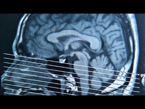
Magnetic resonance imaging (MRI) scans are mainly used to diagnose cavernomas. As symptoms aren’t always evident, many people are only diagnosed with a cavernoma after having an MRI scan for another reason.

computerised tomography (CT) scan or angiography can also be used to diagnose cavernoma, but they’re not as reliable as an MRI scan.

Monitoring your symptoms

Any symptoms you have may come and go as the cavernoma bleeds and then reabsorbs blood. It’s important to closely monitor your symptoms, as any new symptoms might be a sign of a haemorrhage.

Your doctor can advise you about what to do if you experience any new or worsening symptoms. They may also recommend having a further brain scan.

MRI and CT scans can be used to detect bleeding on the brain, although they can’t necessarily identify cavernomas that are at an increased risk of bleeding.

This is because the features of a cavernoma that can be seen on a brain scan, such as an increase in size, don’t appear to be directly linked to the likelihood of bleeding.

Although cavernomas can get bigger, large cavernomas aren’t any more likely to bleed than smaller ones.

What are the chances of a cavernoma bleeding?

The risk of having a haemorrhage varies from person to person, depending on whether you have experienced any bleeding before.

If you haven’t had any bleeding before, it’s estimated you have a less than 1% chance of experiencing a haemorrhage each year.

If your cavernoma has bled previously, your risk of having another haemorrhage is somewhere between 4% and 25% each year.

However, this risk decreases progressively over time if you don’t experience any further bleeds, and eventually returns to the same level as that of people who haven’t had any bleeding before.

Your level of risk will be one of the main factors taken into consideration when deciding if you would benefit from treatment.

Treating cavernoma

The recommended treatment for cavernoma will vary depending on an individual’s circumstances and factors such as size, location and number.

Some cavernoma symptoms, such as headaches and seizures, can be controlled with medication.

However, more invasive treatment may sometimes be offered to reduce the risk of future haemorrhages. The decision to have such treatment is made on a case-by-case basis in discussion with your doctor.

Types of treatment offered in the UK to reduce the risk of haemorrhages include:

neurosurgery – carried out under general anaesthetic to remove the cavernoma

stereotactic radiosurgery – where a single, concentrated dose of radiation is aimed directly at the cavernoma, causing it to become thickened and scarred

In most cases, neurosurgery is preferred to stereotactic radiosurgery because the effectiveness of radiosurgery in preventing haemorrhages is unknown.

Stereotactic radiosurgery is usually only considered if the position of the cavernoma makes neurosurgery too difficult or dangerous.

Risks of invasive treatment include stroke and death, although the exact risks depend on the location of the cavernoma. You should discuss the possible risks of treatment with your doctor beforehand.

Further information

International research programmes are trying to find out more about what causes cavernoma and how these defective blood vessels are formed. The long-term outlook for people with cavernomas is also being investigated.

The Cavernoma Alliance UK website has more information about the condition.
Von Willebrand disease – what are the causes and signs of Von Willebrand disease?

Von Willebrand Disease

Von Willebrand Disease

Introduction

Von Willebrand disease is the most common inherited bleeding disorder.

People with Von Willebrand disease (VWD) either have a deficiency of a blood protein called Von Willebrand factor, or this protein doesn’t work properly. This means that their blood cannot clot properly and they are prone to prolonged or excessive bleeding.

Von Willebrand factor acts like a glue which allows platelets (a type of blood cell) to stick together and to plug up damaged blood vessels. It also carries and protects another clotting factor called FVIII (factor 8).

Signs and symptoms

VWD can cause problems at any time from birth, but for many people the symptoms are so mild that they may never know they have it.

It’s estimated that around 1% of the UK population have reduced levels of Von Willebrand factor, but only a small minority have symptoms that might require medical attention.

Symptoms of VWD can include:

bruising easily

bleeding from the gums, nose and lining of the gut

prolonged bleeding from cuts

excessive bleeding after tooth extraction or surgery

in women, heavy periods

With treatment, even people with more severe symptoms can go on to lead normal and active lives.

What causes VWD?

VWD is usually caused by a fault in the gene responsible for the production of Von Willebrand factor. This genetic fault can be passed on to a child by one or both of their parents.

There are four different types of VWD. The condition is classified according to the amount and function of the Von Willebrand factor in the blood.

The main types are:

Type 1 – The mildest and most common type. People with type 1 VWD have a reduced level of Von Willebrand factor in their blood. Symptoms might be so mild they never know they have VWD. They don’t usually bleed spontaneously, but can bleed significantly if they have surgery, injure themselves or have a tooth removed.

Type 2 – In this type, which can itself be divided into four further subtypes, the Von Willebrand factor doesn’t work properly. The symptoms of type 2 VWD are often more severe than type 1, but milder than type 3.

Type 3 – People with this rare type have very low levels of Von Willebrand factor in their blood, or none at all. This causes problems similar to haemophilia. Bleeding from the mouth, nose and bowel from an early age is typical, and joint and muscle bleeds can occur after injury.

Pseudo, or platelet type – this is similar to type 2, but instead of the abnormality occurring in the Von Willebrand factor, it occurs in the platelets.

Types 1, 2 and pseudo-VWD are often passed on through “autosomal dominant inheritance”. This means that only one parent has to pass a copy of the faulty gene to their child for them to develop the condition.

Type 3 VWD is passed on through “autosomal recessive inheritance”. This means the child needs to inherit a copy of the faulty gene from both parents. In such cases, the parents may have no symptoms themselves because they may have only inherited a copy of the faulty gene from one of their own parents, rather than both.

Read more about genetic inheritance.

Diagnosing VWD

The symptoms of VWD can be mild and this can mean it is sometimes difficult to diagnose.

If a doctor suspects you may have VWD, you should be referred to a haematologist (a specialist in diagnosing and treating blood disorders) for tests and any necessary treatment.

A blood test can usually be carried out to diagnose the condition, although this may occasionally have to be repeated because levels of Von Willebrand factor can vary over time. You will also be asked about your family history to check if any other members of the family have a bleeding disorder.

Managing VWD

Mild VWD might not need any treatment, and you may be able to manage the condition by taking special precautions and making a few lifestyle changes.

Otherwise, three main treatments are available to either prevent or treat bleeding:

desmopressin – available as a nasal spray or injection

tranexamic acid – available as tablets, a mouthwash or an injection

Von Willebrand factor concentrate – this is purified from human plasma (from donated blood) and is given directly into a vein

Your doctor will explain which medication is suitable for you and when you should take it.

Your doctor will often recommend a “test dose” of desmopressin to check that it works well. Common side effects include facial flushing, palpitations, changes in blood pressure, and sometimes a mild headache. It can also cause fluid retention for up to 24 hours after it is taken, so your doctor will ask you to restrict the amount of fluid you drink after treatment.

Some advice on the use of these medications is given below, along with general lifestyle changes you can make.

General advice

Most people with VWD have the mildest type, type 1, and may not even realise they have it. They can live normal and active lives and take part in most sports and activities.

In more severe cases, contact sports may need to be avoided. Protective equipment should also be worn for certain activities where there is a risk of bleeding or bruising.

If someone with VWD has an accident or is scheduled for surgery, it is important that the doctor or surgeon is aware the person has the condition.

Due to the risk of excessive bleeding, boys at risk of VWD should not be circumcised without first speaking to a doctor.

Managing heavy periods

Girls and women with VWD who have heavy periods might want to carry extra pads or even a change of clothes in case of excessive bleeding.

They should get advice from their GP or gynaecologist about options to control their bleeding, these include:

the oral contraceptive pill

the intrauterine system (IUS)

tranexamic acid tablets

a desmopressin nasal spray

in severe cases, Von Willebrand factor concentrate

Read more about treating heavy periods.

Managing nosebleeds and other injuries

If bleeding does occur, applying pressure to the area may be all that’s needed. For example, someone who gets a nosebleed should put pressure on the bridge of the nose to help stop the bleeding.

Taking medication continuously to prevent bleeding and bruising is not usually necessary, although your doctor may prescribe desmopressin or tranexamic acid for you to take in the event of an injury or bleed.

Injections of Von Willebrand factor concentrate may be needed to treat injuries in people with type 3 or type 2 VWD, or particularly severe injuries in people with type 1 VWD.

Preventing bleeding before dental procedures or surgery

People with type 1 VWD can usually be treated with desmopressin before dental procedures or surgery.

Desmopressin temporarily boosts levels of Von Willebrand factor long enough to prevent bleeding during the procedure. Tranexamic acid is also often given in addition to desmopressin.

This may not necessarily be helpful in treating people with some forms of type 2 VWD, and is not at all effective in type 3 VWD (as there are no levels of Von Willebrand factor to boost). In these cases, injections of Von Willebrand factor concentrate may be needed instead.

Patients with type 1 also might need Von Willebrand factor concentrate before major surgery, although desmopressin and tranexamic acid usually offer enough protection against bleeding.

Advice for pregnant women

Levels of Von Willebrand factor often increase throughout pregnancy – but despite this, some pregnant women with VWD are at increased risk of bleeding complications during labour and after delivery.

As long as the doctors and midwives are aware of your condition, you can be monitored carefully and given treatment, if required, to prevent bleeding during and after delivery.

Factor V Leiden – what are the signs and symptoms of Factor V Leiden?

Factor V Leiden

Factor V Leiden

Introduction

Thrombophilia is where the blood has an increased tendency to form clots.

It’s not a condition or a disorder itself, and is sometimes associated with blood clots (venous thromboses) that are caused by genetic changes (mutations).

If you have thrombophilia, you’re more likely to develop a blood clot in one of the large veins in your leg. This is known as deep vein thrombosis (DVT).

It’s important to be aware of the signs of a DVT, such as unexplained pain, swelling or tenderness in your leg (usually in your calf). See your GP as soon as possible if you have these symptoms (see below).

The blood clotting process

When you cut yourself, the bleeding usually triggers a number of chemical reactions in the blood.

The chemical reactions cause a blood clot to form, which sticks to the injured part of the blood vessel along with blood cells called platelets.

Clotting substances, known as clotting factors, help control bleeding and work with platelets to ensure the blood clots effectively.

However, in thrombophilia, there’s an imbalance in clotting chemicals and a person with the disorder either has too much clotting factor, or too little of the substance that prevents clotting.

Types of thrombophilia

There are many different types of thrombophilia. Some of the main types are outlined below.

Factor V Leiden

Factor V Leiden is a type of thrombophilia caused by a specific gene mutation.

It’s the most common type of inherited thrombophilia, with 3-8% of Europeans having one copy of the factor V Leiden mutation in each cell, and about one in 5,000 people having two copies of the mutation. It’s less common in other populations.

If you have Factor V Leiden, your risk of developing a DVT is increased (see above). There’s also a higher risk of blood clots breaking away from their original location and travelling to other areas of your body through your bloodstream.

This can be particularly serious if the blood clot becomes stuck in the main artery of your lungs (the pulmonary artery). This is known as a pulmonary embolism and it can be life-threatening (see below).

Prothrombin 20210 mutation

Prothrombin 20210 mutation, also known as Factor II mutation, is another genetic blood disorder that increases the risk of clots.

Prothrombin is a protein in the blood which helps it to clot (thicken). However, in some people, a mutation in the gene that produces prothrombin causes too much to be produced, resulting in an increased tendency for blood clots, such as DVTs, to form.

As with Factor V Leiden, prothrombin 20210 is more common in white people, particularly Europeans.

Protein C, protein S or antithrombin deficiency

Protein C, protein S, and antithrombin are natural anticoagulants (substances that prevent the blood from clotting).

If you have low levels of these anticoagulants, or if they don’t work properly, your risk of developing a DVT or pulmonary embolism is increased.

Problems with protein C, protein S or antithrombin can be inherited but are rare.

Antiphospholipid syndrome

Antiphospholipid syndrome, also known as Hughes syndrome, is an immune system disorder that increases the risk of blood clots.

Your body produces antibodies that attack phospholipids, a fat molecule thought to keep blood at the right consistency.

The antibodies bind to the phospholipids, increasing your risk of a blood clot developing in a vein or artery.

Symptoms of thrombophilia

Thrombophilia is often mild and many people don’t experience any health problems. Symptoms will only occur if the disorder causes a blood clot.

People with thrombophilia are particularly at risk of developing a DVT.

Warning signs of a DVT include:

pain, swelling and tenderness in one of your legs (usually your calf)

a heavy ache in the affected area

warm skin in the area of the clot

redness of your skin, particularly at the back of your leg below the knee

DVT usually, although not always, affects one leg. The pain may be worse when you bend your foot up towards your knee.

Part of the blood clot can sometimes break away and travel through the bloodstream. This is known as an embolism.

Embolisms can be dangerous if they become lodged in the lungs (pulmonary embolism). This is a serious and potentially life-threatening condition because it can prevent blood from reaching your lungs.

The symptoms of a pulmonary embolism are:

chest or upper back pain

shortness of breath

coughing – usually dry but you may cough up blood or mucus containing blood

feeling lightheaded or dizzy

fainting

See your GP immediately if you experience any combination of the above symptoms. Alternatively, you can call NHS 111 or contact your local out-of-hours service for advice. Dial 999 to request an ambulance if your symptoms are severe.

Diagnosing thrombophilia

Thrombophilia testing isn’t routinely carried out on everyone who has a blood clot.

Testing for inherited thrombophilia in people with venous thrombosis isn’t usually needed because the results don’t usually influence clinical decisions.

For example, decisions regarding the length of treatment should be made by taking into consideration whether or not venous thrombosis was provoked, whether other risk factors are present, and the risk of bleeding due to treatment with anticoagulants, regardless of whether inherited thrombophilia is present or not.

Thrombophilia is diagnosed by having blood tests weeks or months after your blood clot. The tests look for anticoagulant deficiencies. Before having the tests, you’ll usually have to wait until you’ve stopped taking anticoagulant medicine, such as warfarin, for four to six weeks.

If the blood test results indicate you have thrombophilia, you may be referred to a haematologist (a specialist in diagnosing and treating blood disorders).

Treating thrombophilia

Treatment may not be needed if you have mild thrombophilia.

If you develop a blood clot, you’ll need treatment for the blood clot and treatment to prevent further clots. You may need to take warfarin tablets or have an injection of heparin.

Warfarin and heparin

Warfarin and heparin are anticoagulants . They interfere with the clotting process and are commonly used to treat or prevent DVT and pulmonary embolisms.

If you need an anticoagulant to prevent a clot, you’ll be prescribed warfarin. It will take a few days to work properly.

If you need instant treatment for an existing clot, you’ll usually be given a heparin injection (which works straight away), as well as warfarin tablets for the first few days. The injection will either be given in hospital or at home.

A heparin injection may also be given to people with antiphospholipid syndrome, before and after surgery or during pregnancy. Unlike warfarin, heparin is safe to take in pregnancy.

International Normalised Ratio (INR) test

Your doctor will need to adjust your warfarin dose so your blood doesn’t clot easily, but isn’t too high to put you at risk of bleeding problems.

You’ll need to have a regular blood test called the International Normalised Ratio (INR) to measure your blood clotting ability while taking warfarin.

You’ll have the INR test less frequently once your ideal dose has been reached (an INR of two to three is usually the aim).

Lifestyle advice

If you have thrombophilia, it’s important you’re aware of the symptoms of a blood clot (see above), and that you see your GP immediately if you think you have one.

You should also take the following precautions to lower your risk of blood clots:

lose weight if you’re overweight

stop smoking if you smoke

eat a healthy, balanced diet and exercise regularly

avoid being immobile for long periods because this can cause a DVT (read more about preventing DVT)

If you’re pregnant, or planning to get pregnant, discuss this with your GP and inform the midwife and obstetrician about your condition.

You may need to take low-dose aspirin or an anticoagulant medicine while you’re pregnant to prevent problems occurring during pregnancy or miscarriage.

If you’re having a major operation, make sure you tell the healthcare professionals treating you about your condition. You may need a heparin injection to prevent a blood clot forming.

Women with thrombophilia should avoid taking the combined oral contraceptive pill or hormone replacement therapy (HRT) because they’ll further increase your risk of developing a blood clot.

World Sepsis Day – Tuesday 13 September – What are the signs and symptoms of sepsis?

World Sepsis Day 2016

World Sepsis Day 2016

What are the signs and treatment of sepsis?

“The figures are huge” says John Parker who is Leicester’s Hospitals Sepsis Lead Consultant , “every year in the UK there are 150,000 cases of Sepsis, resulting in a staggering 44,000 deaths – more than bowel, breast and prostate cancer combined. Unfortunately the initial signs are often ignored, an individual may say “I just felt like I had a bout of flu, stomach upset or chest infection” and so people don’t get any medical attention. Early diagnosis is essential, so we want to highlight the signs of Sepsis and raise its awareness.”

“It’s important to remember that sepsis is a medical emergency. Every minute and hour counts, especially since the infection can spread quickly. There’s no one symptom of sepsis, but rather it has a combination of symptoms.”

Sepsis is caused by the way the body responds to germs, such as bacteria, getting into your body. The infection may have started anywhere in a sufferer’s body, and may be only in one part of the body or it may be widespread. Sepsis can occur following chest or water infections, problems in the abdomen like burst ulcers, or simple skin injuries like cuts and bites.

“A lot of people will more readily know sepsis as septicaemia or blood poisoning and whilst diagnosis and treatment can be different for adult and children, the basic signs to look out for are:

S lurred speech
E xtreme shivering or muscle pain
P assing no urine (in a day)
S evere breathlessness
I feel like I might die
S kin mottled or discoloured

Amandeep Sadhra who has recovered from sepsis says “I was just going about my normal day, when I noticed a rash on my hand, I didn’t really take any notice of it as I suffer from eczema, but during the course of the day it got worse and was throbbing a lot. By the time I got home after work, I felt very tired and had no appetite. I decided to just take some paracetamol and go to bed. The next day I felt no better and didn’t want to get up and my hand had ballooned up like a boxing glove and I was starting to shiver, I felt like I was getting a fever. It was at this time my husband said we should go to the Emergency Department.

“I received scans, a blood test and was advised that as there was a lot of fluid on my hand that I would have to have an operation, but during the course of the night the doctor advised me that my blood pressure was dropping and the antibiotics were not working and I was going to be transferred to intensive care. The next day I was taken for my operation and woke up five or so days later after my procedure. I was then advised that I had been very ill after my operation, suffering from multiple organ failure, slight pneumonia and it was decided to continue my sedation. I was then advised that I had Sepsis.”

“It was a life-changing event, I had always been fit and healthy but after being discharged from hospital it has taken me several months to recuperate, go back to work, get back to normal. You never think something like this could happen to you, particularly from something so minimal like a skin rash to something life threatening.”

If you suspect sepsis, get medical attention immediately.

Blood Donation: The Easy Way to Save Lives

It seems hard to believe that a single unit of blood can save up to three people’s lives, but that is the truth. By giving up this small sample of our blood, along with little more than an hour of our time, we could possible give three people a second chance in life. One of those could be a family member or a friend who is urgently in need of a donation. A small sacrifice for you is quite possibly a life-saving intervention for them.

Blood Donation - The Easy Way to Save Lives

Blood Donation – The Easy Way to Save Lives

Deciding to give blood isn’t as simple as wanting to give it, though. A number of factors could deem your blood unsuitable for donation, such as any recent pregnancy or childbirth, or undergoing a major operation inside the last six months. While wanting to donate blood is admirable, you should only make a donation if you are sure that your blood would be safe to give to another person. This process can be further complicated by the requirement for compatibility between donor and recipient. People of type A blood, for instance, will not be able to receive from anyone with blood types B or AB, as the presence of the B antigen would cause an antibody against the A antigen and potentially lead to a fatal reaction.

This infographic from Union Quay Medical Centre (http://www.unionquaymedicalcentre.ie/general-practice.html) in Ireland tells you everything you need to know before donating blood for the first time, such as the compatibility of blood groups, the eligibility of donation and the before, during and after of the actual donation process. If you’re thinking of becoming a first-time donor but you have unanswered questions, the guide below could address those uncertainties, so why not take a few minutes to read through it?